You Might Like
-
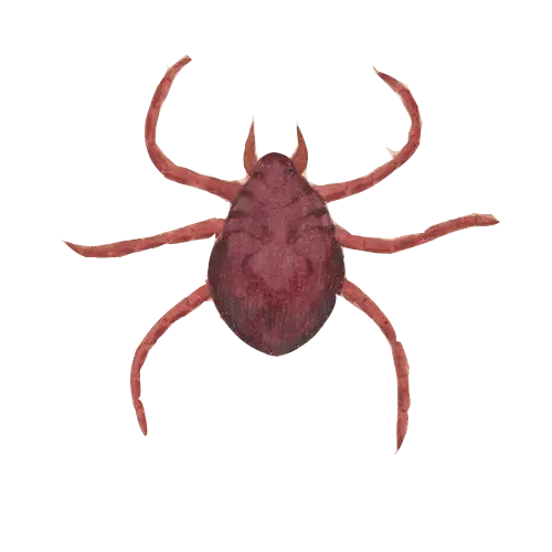
Brown Tick Illustration -

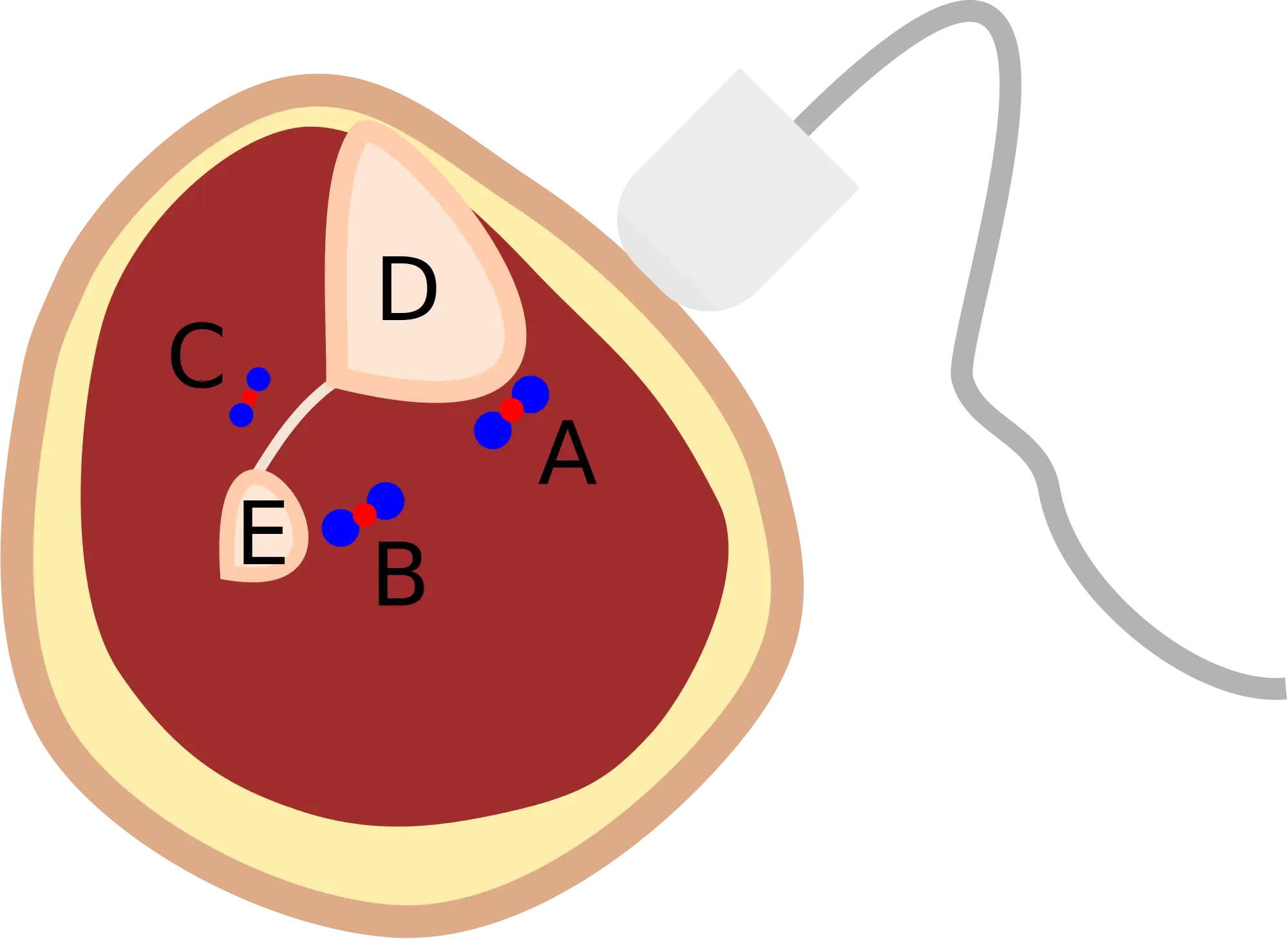
Red Virus Illustration Representing Pathogen -
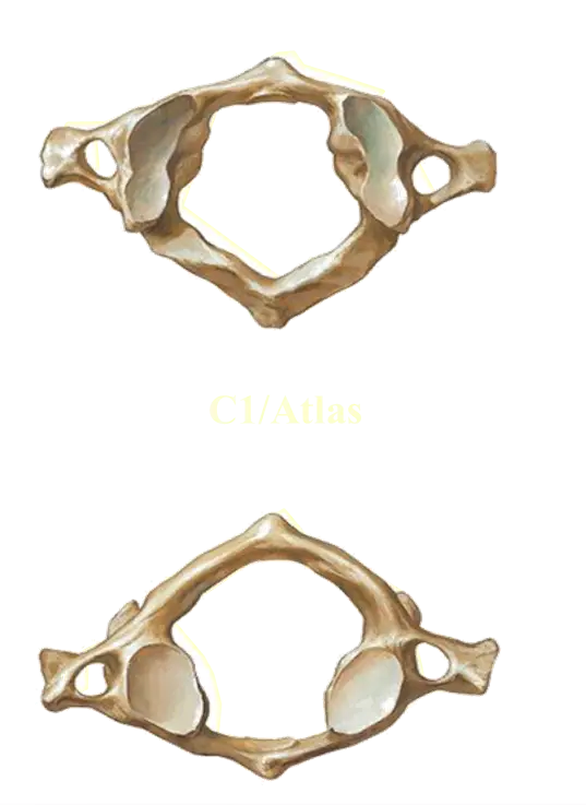
Atlas Vertebra Anatomy -
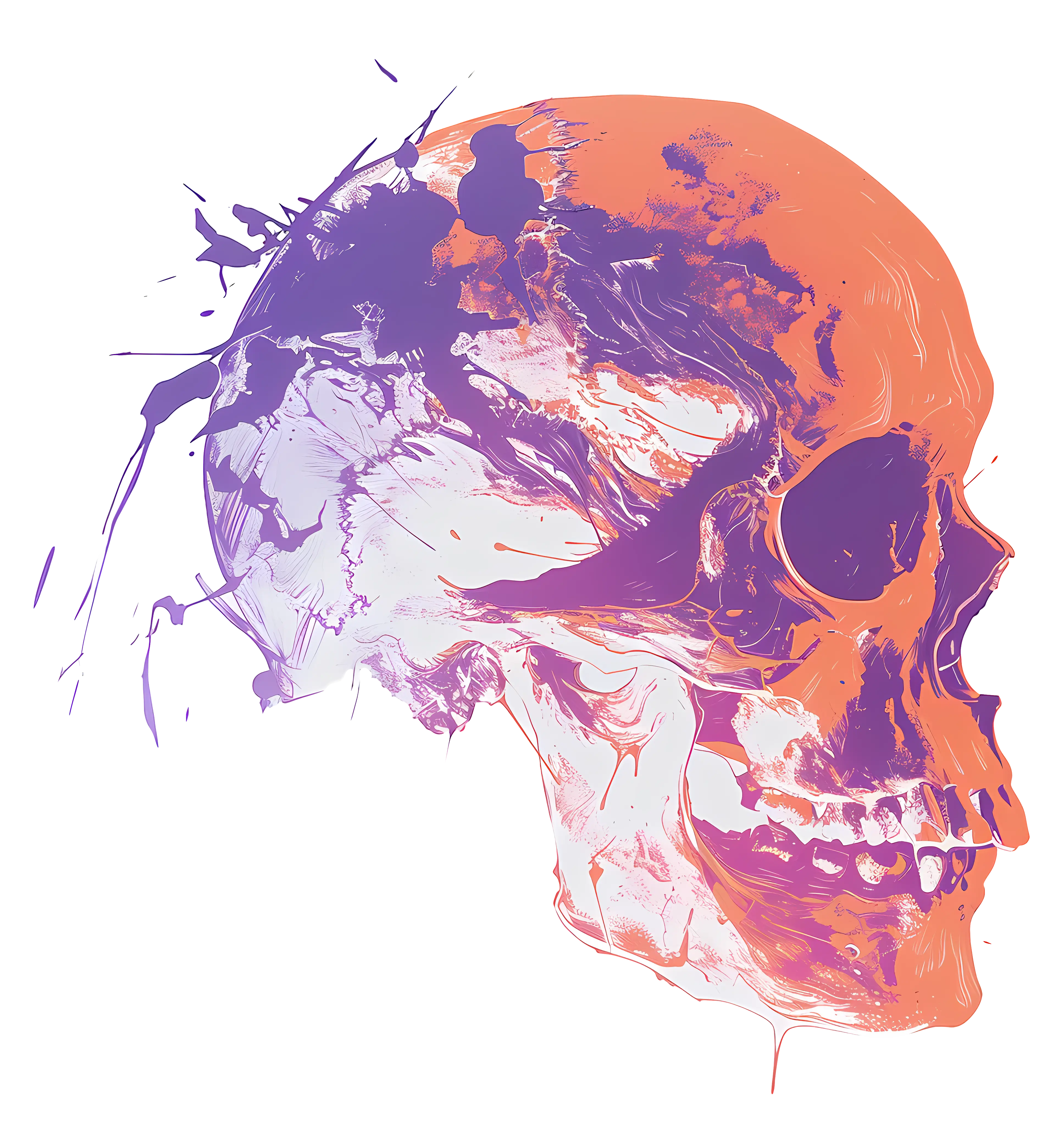
Abstract Colorful Skull Illustration -
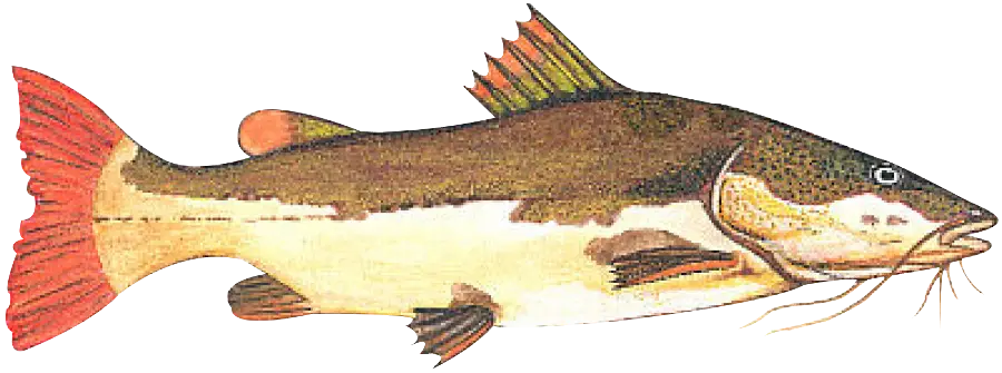
Illustration of a Fish -

Strong Muscular Man Illustration -
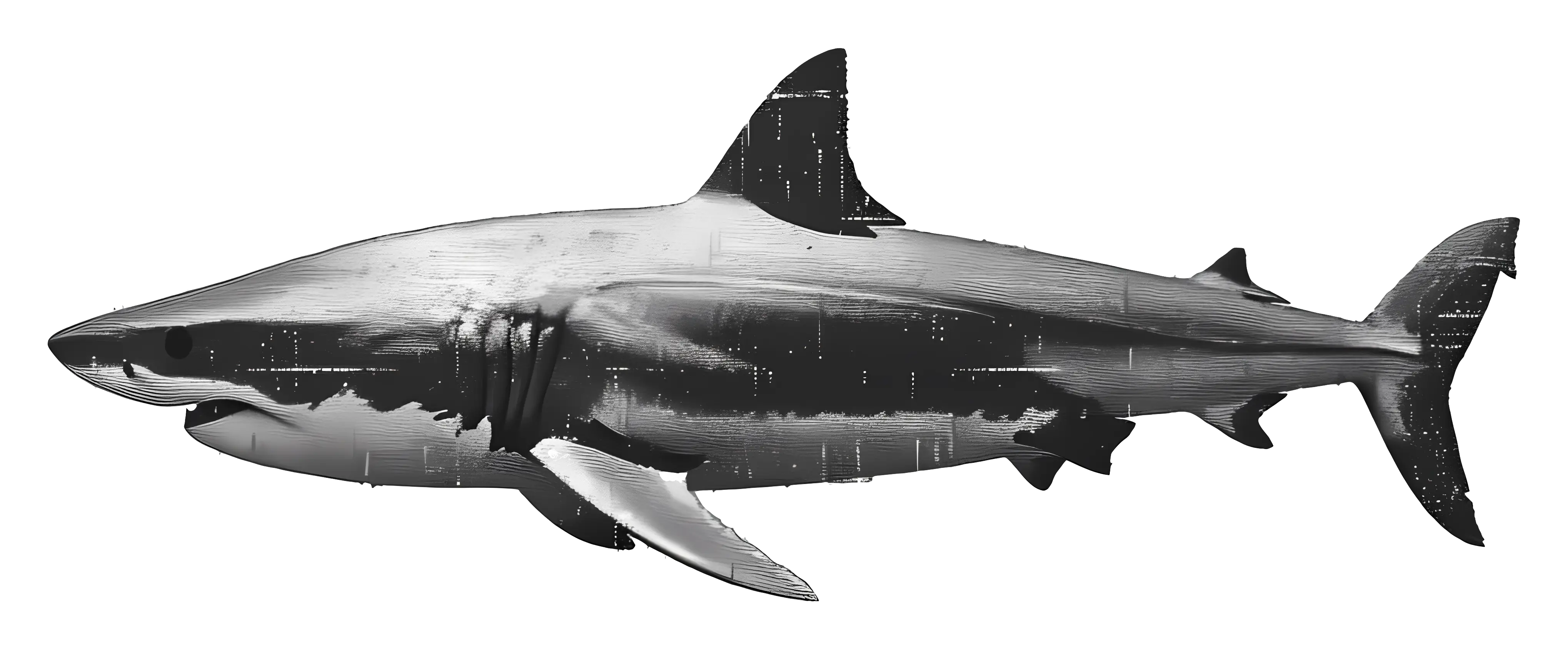
Realistic Shark Illustration -

Cross Made of DNA Strands -
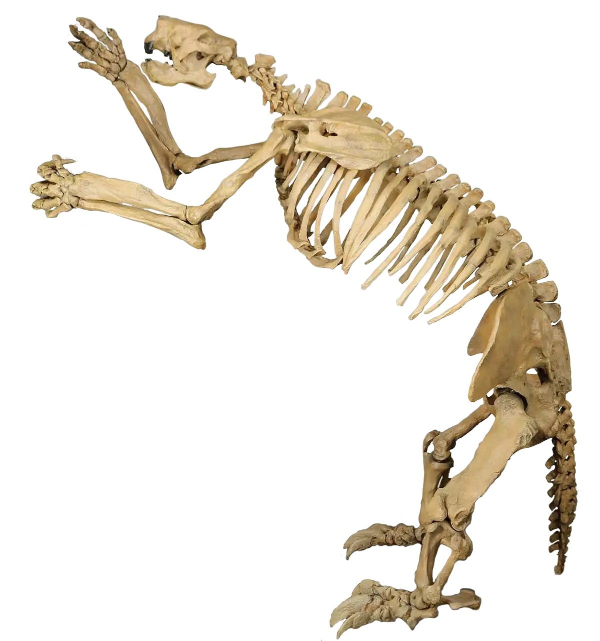
Animal Skeleton Display -
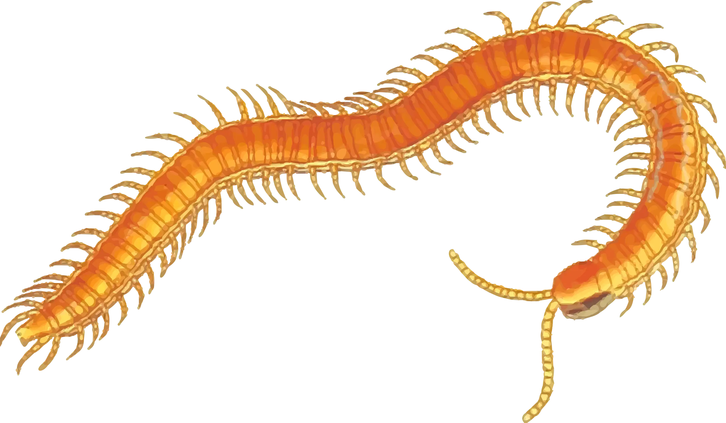
Orange Centipede Illustration -

Blue Human Brain Illustration -
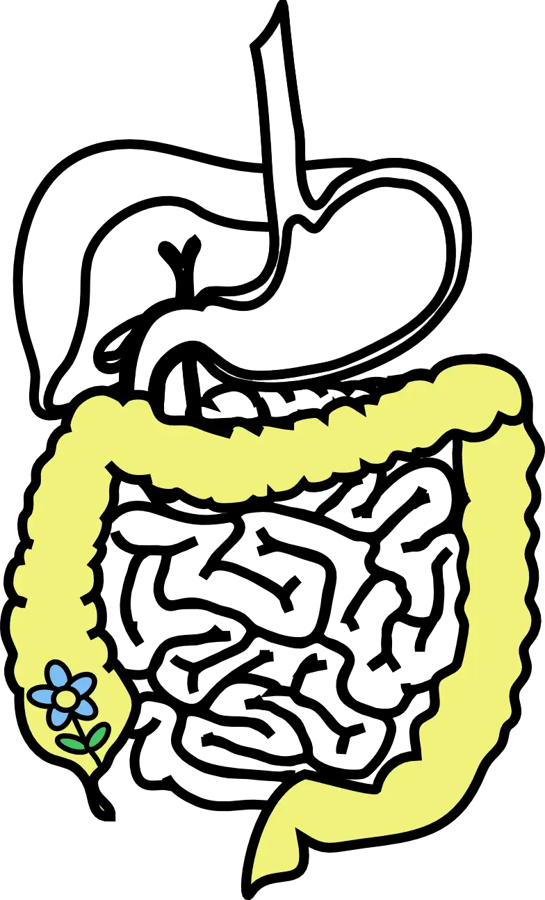
Digestive System Diagram -

HR Workflow Diagram -

Marble Brain Illustration for Education -
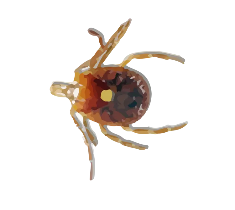
Brown Tick Close-Up Illustration -
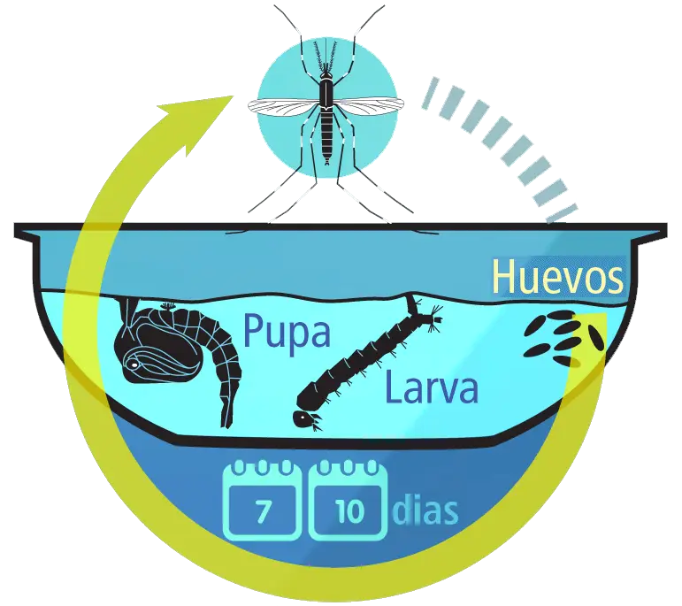
Mosquito Life Cycle Diagram -

Silhouette of Black Hand in Vector Style -
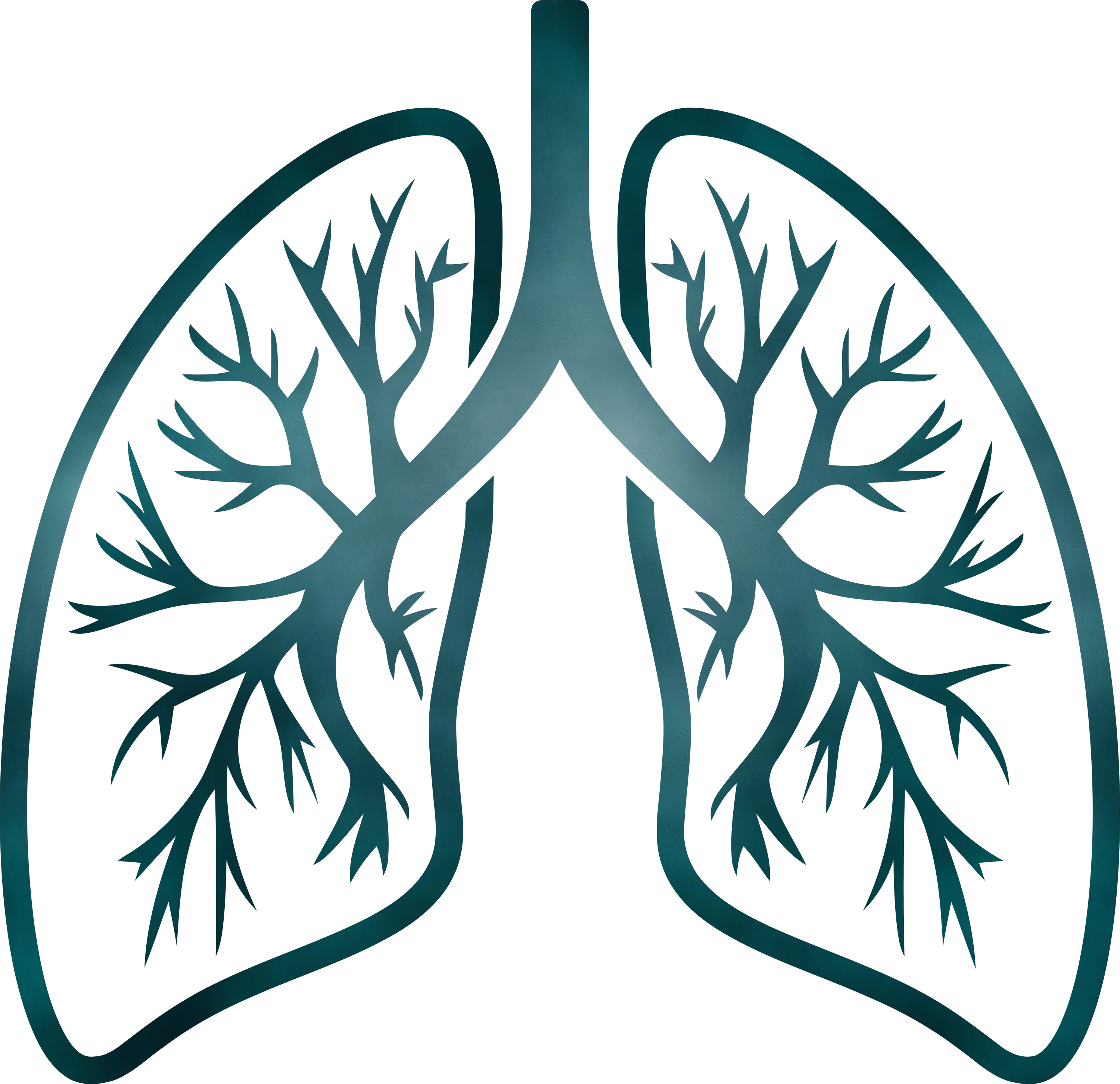
Detailed Lungs Diagram Illustration -

Triangle Diagram for Geometric Representation -
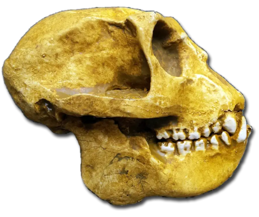
Fossilized Anthropoid Skull Representation -
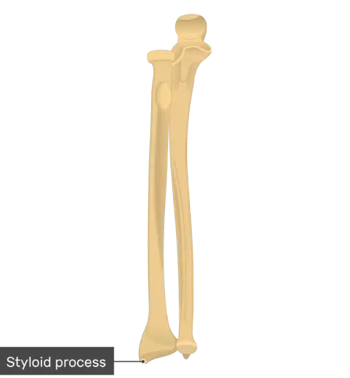
Styloid Process Anatomy Illustration -

Metallic Human Skull Anatomy -
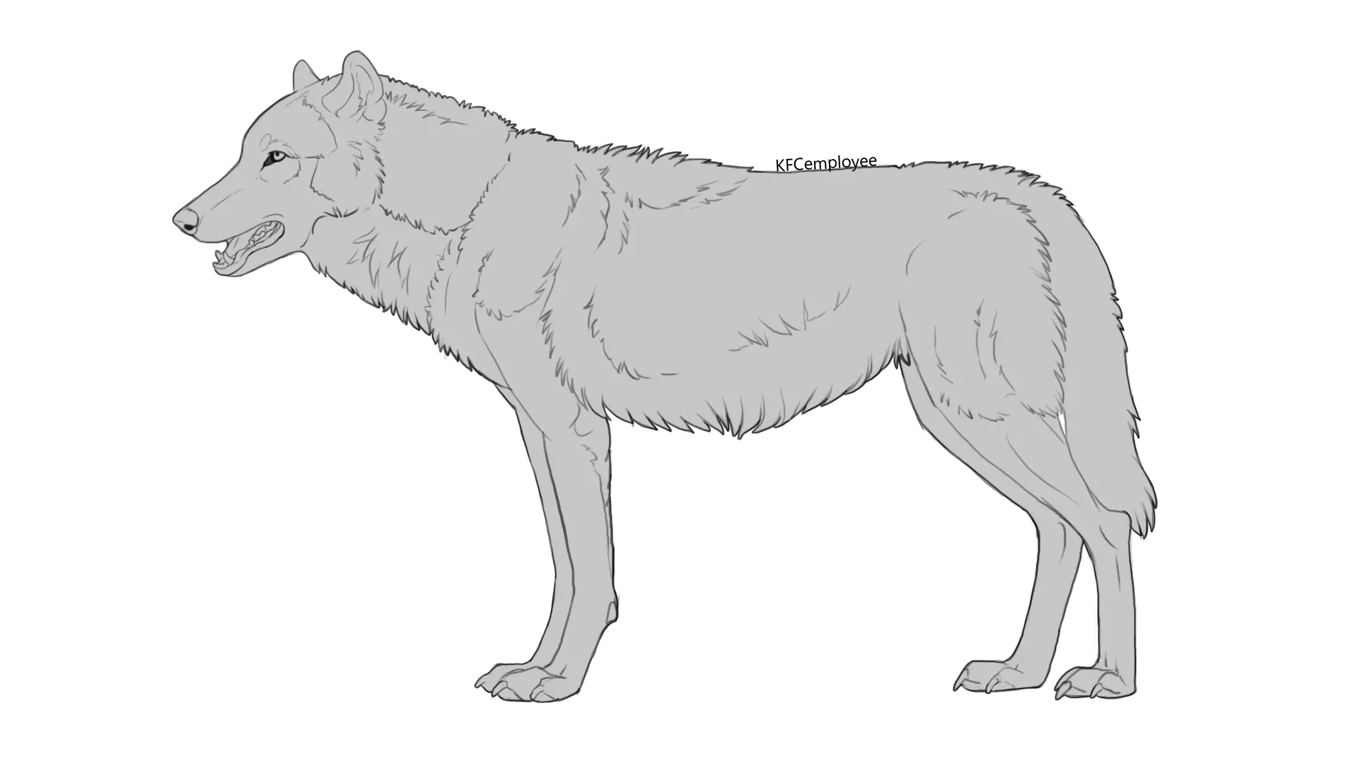
Wolf Outline Illustration -

Detailed Cannabis Plant Illustration -

Black and White Fish Illustration -
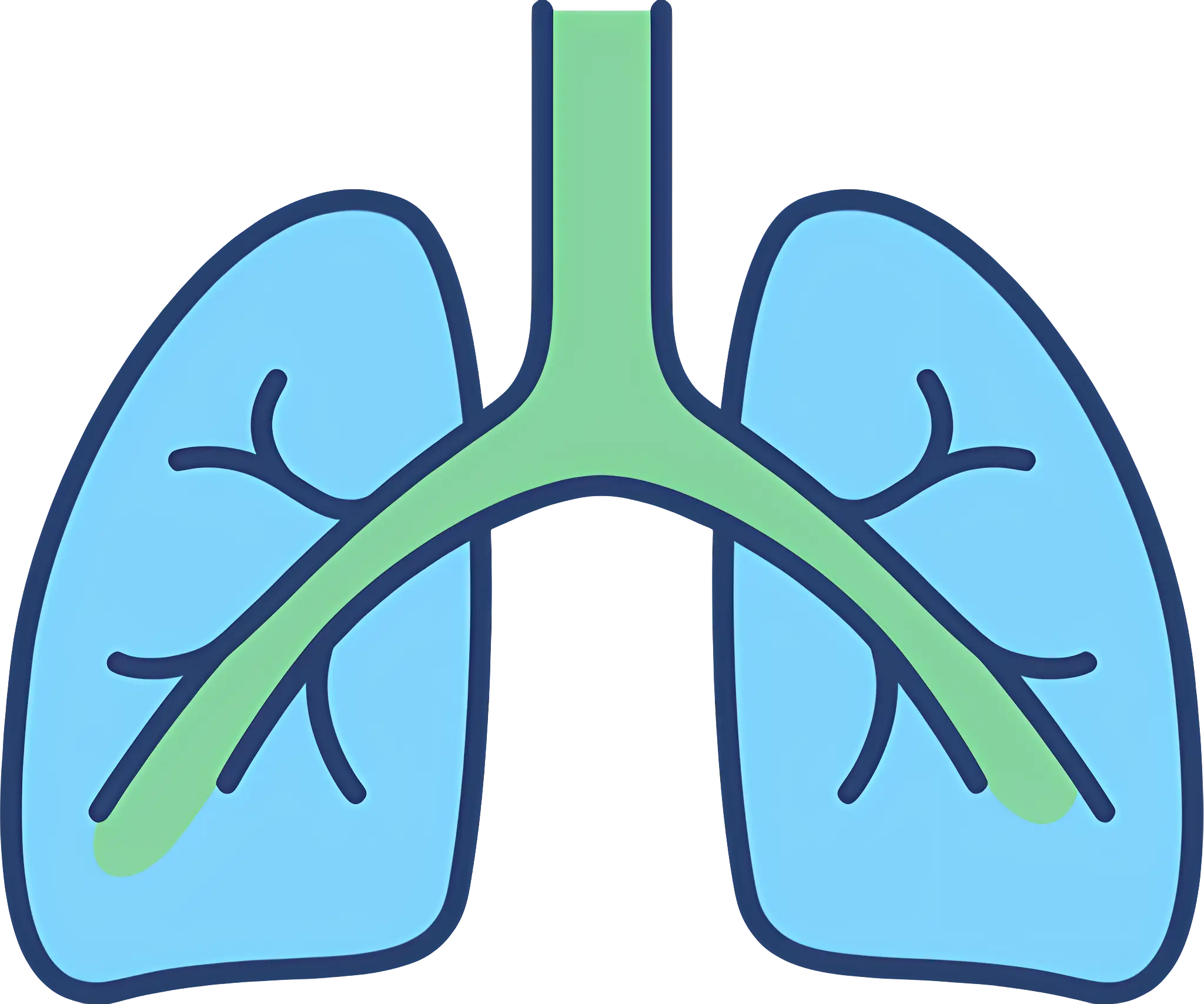
Medical Illustration of Human Lungs -

Mind Map Diagram in Colorful Design -

Green Flowchart Diagram -

Protein Molecular Structure Model -

Mathematical Network Diagram with Nodes and Lines