You Might Like
-

Shiny Red Heart for Valentine's Day -

Red Heart with Valentine's Day Message -

Yellow Flower Heart Arrangement -
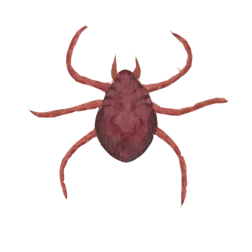
Brown Tick Illustration -
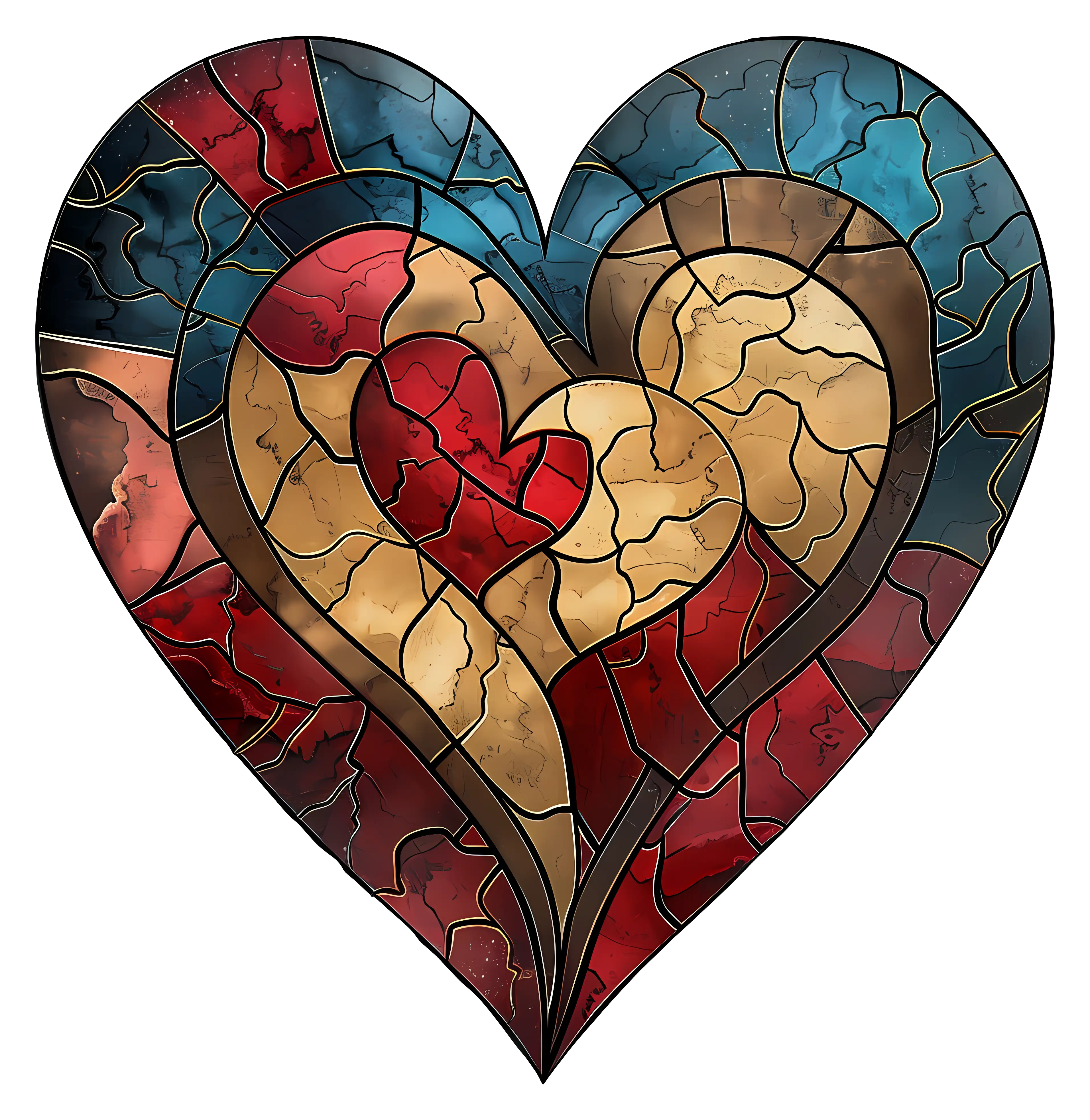
Colorful Stained Glass Heart -

Romantic Swans Forming a Heart -
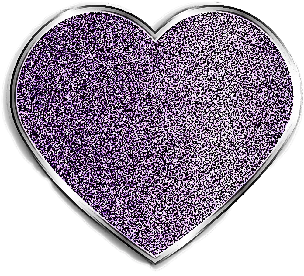
Glittery Purple Heart Symbol -

Heart-Shaped Red Rose Floral Frame -
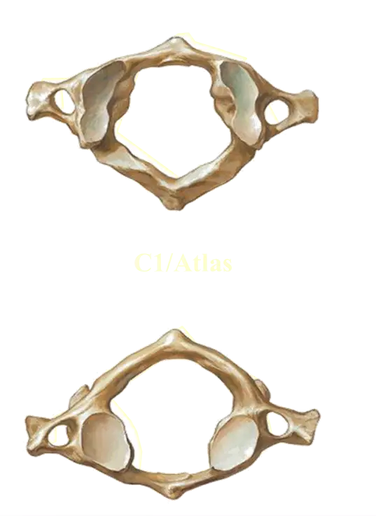
Atlas Vertebra Anatomy -
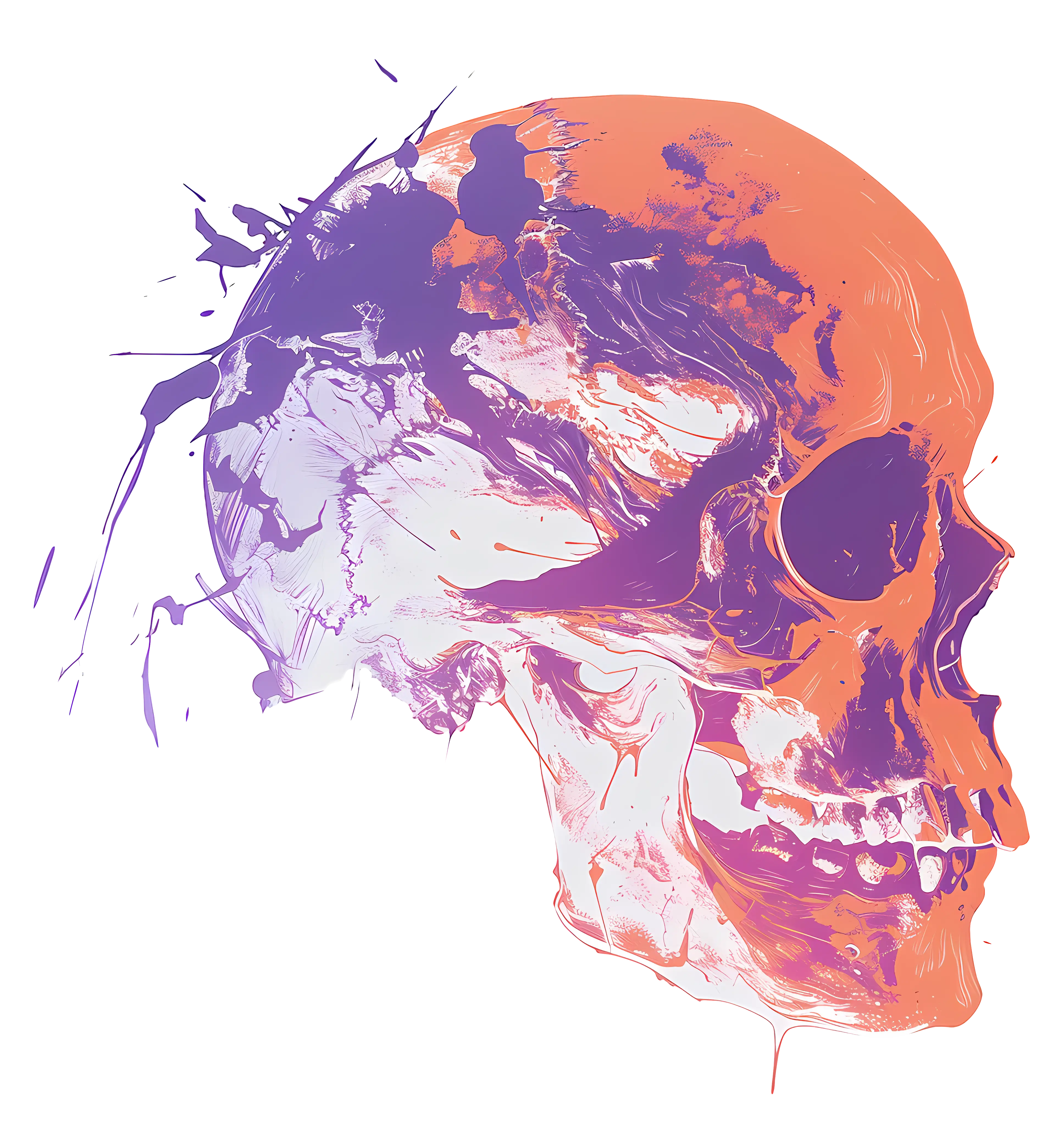
Abstract Colorful Skull Illustration -

Pink Heart Outline Love Symbol -
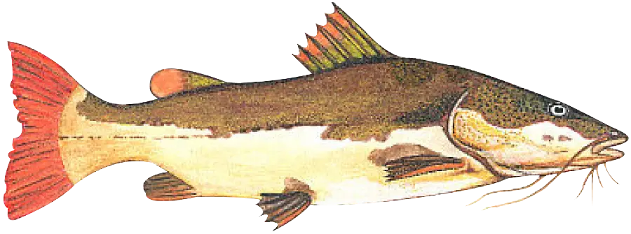
Illustration of a Fish -

Strong Muscular Man Illustration -

Red Heart-Shaped Candy Box Illustration -
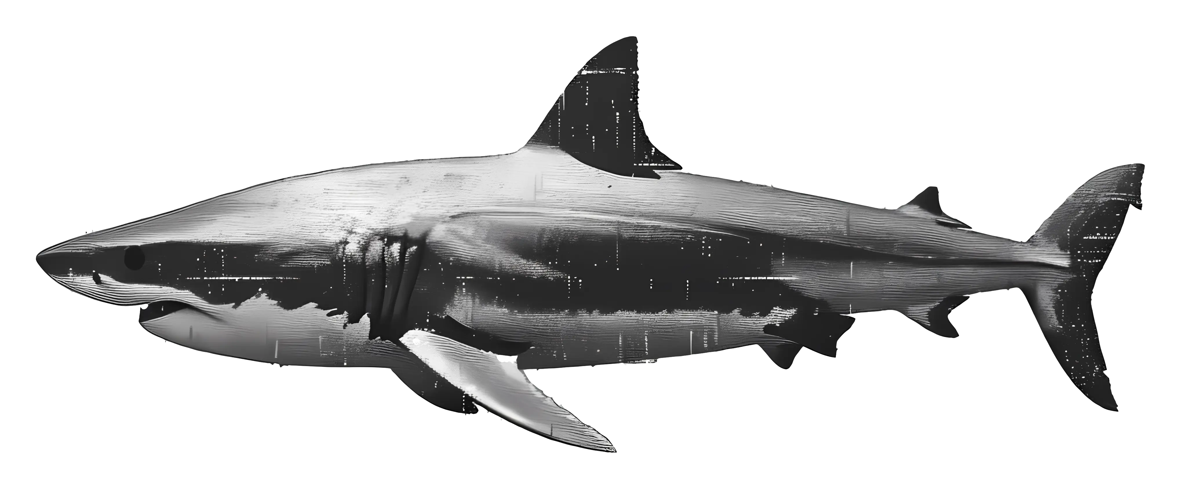
Realistic Shark Illustration -

Heart Shape Made of Pink Leaves -

Rainbow Gradient Heart Illustration -

Red Book with Heart Illustration Design -
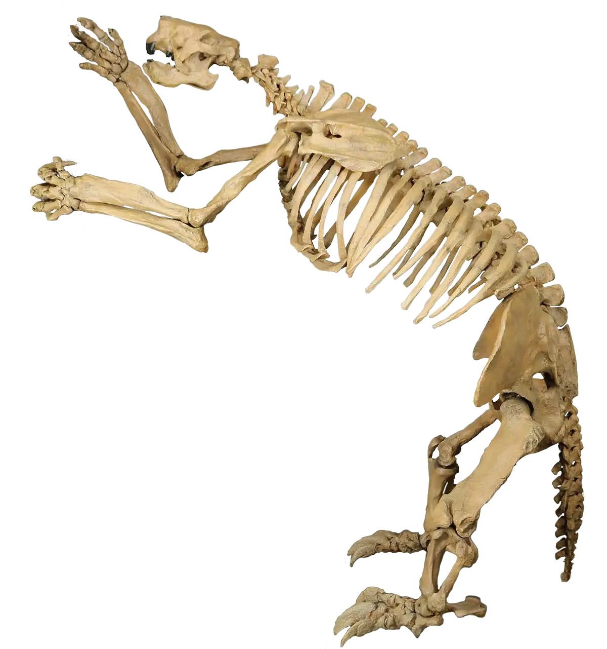
Animal Skeleton Display -

Black Magnifying Glass with Heart Icon -
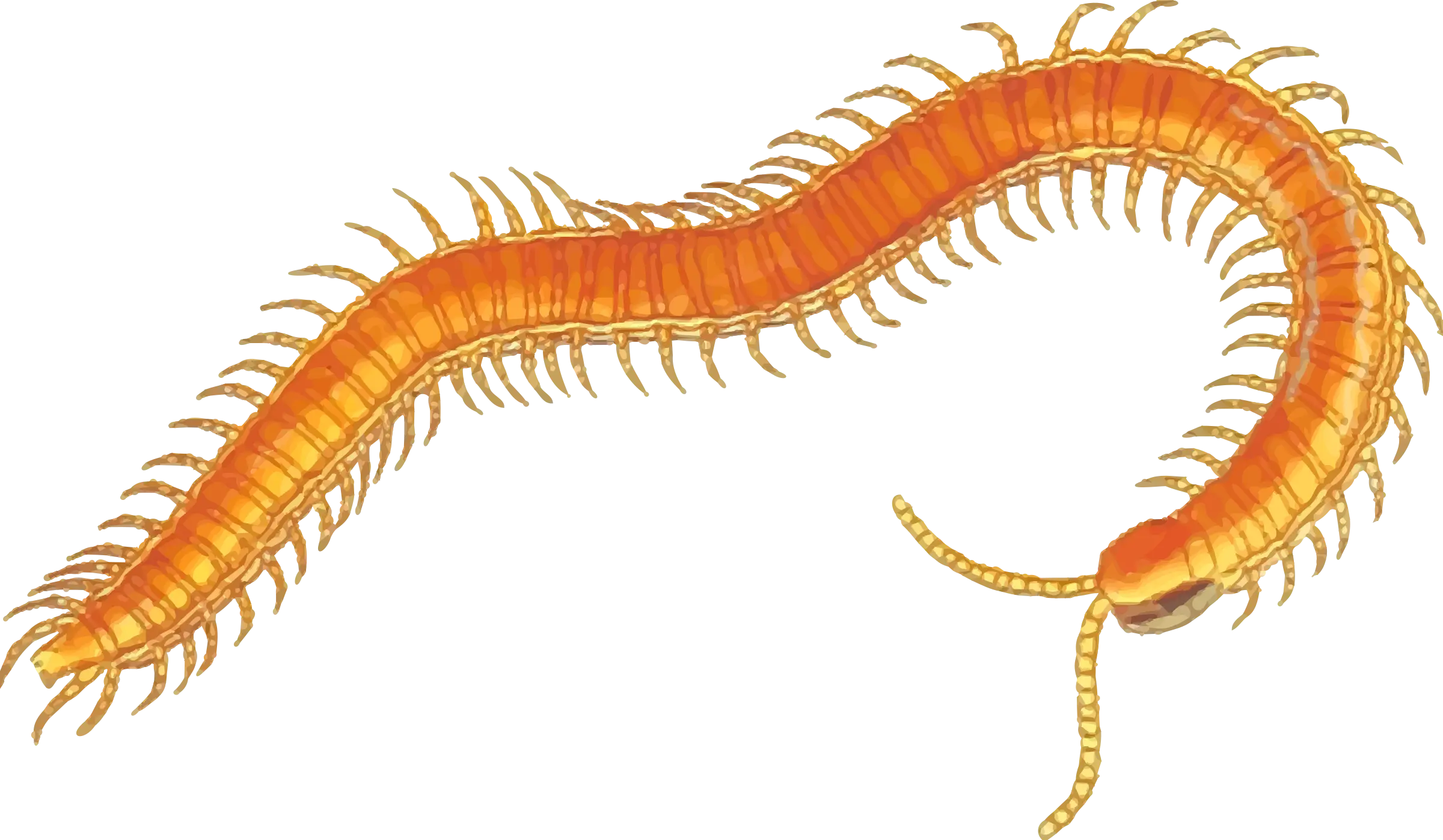
Orange Centipede Illustration -

Fairy Tale Magical Potion Bottle with Florals -
Heart-Shaped German Flag Design -

Stunning Pink Romantic Heart Floral Arrangement -
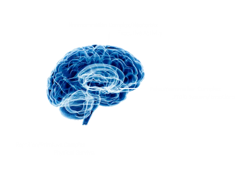
Blue Human Brain Illustration -

Shiny Red Heart Symbol Illustration -
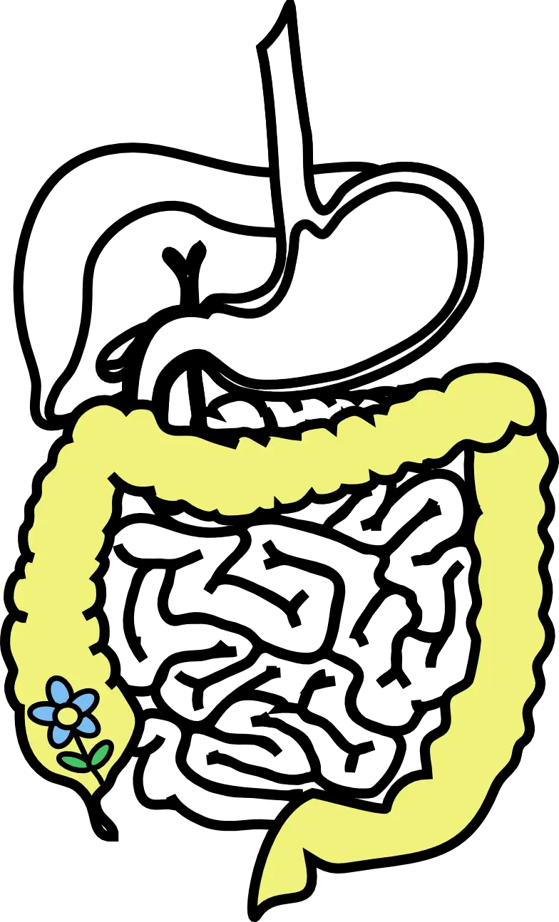
Digestive System Diagram -
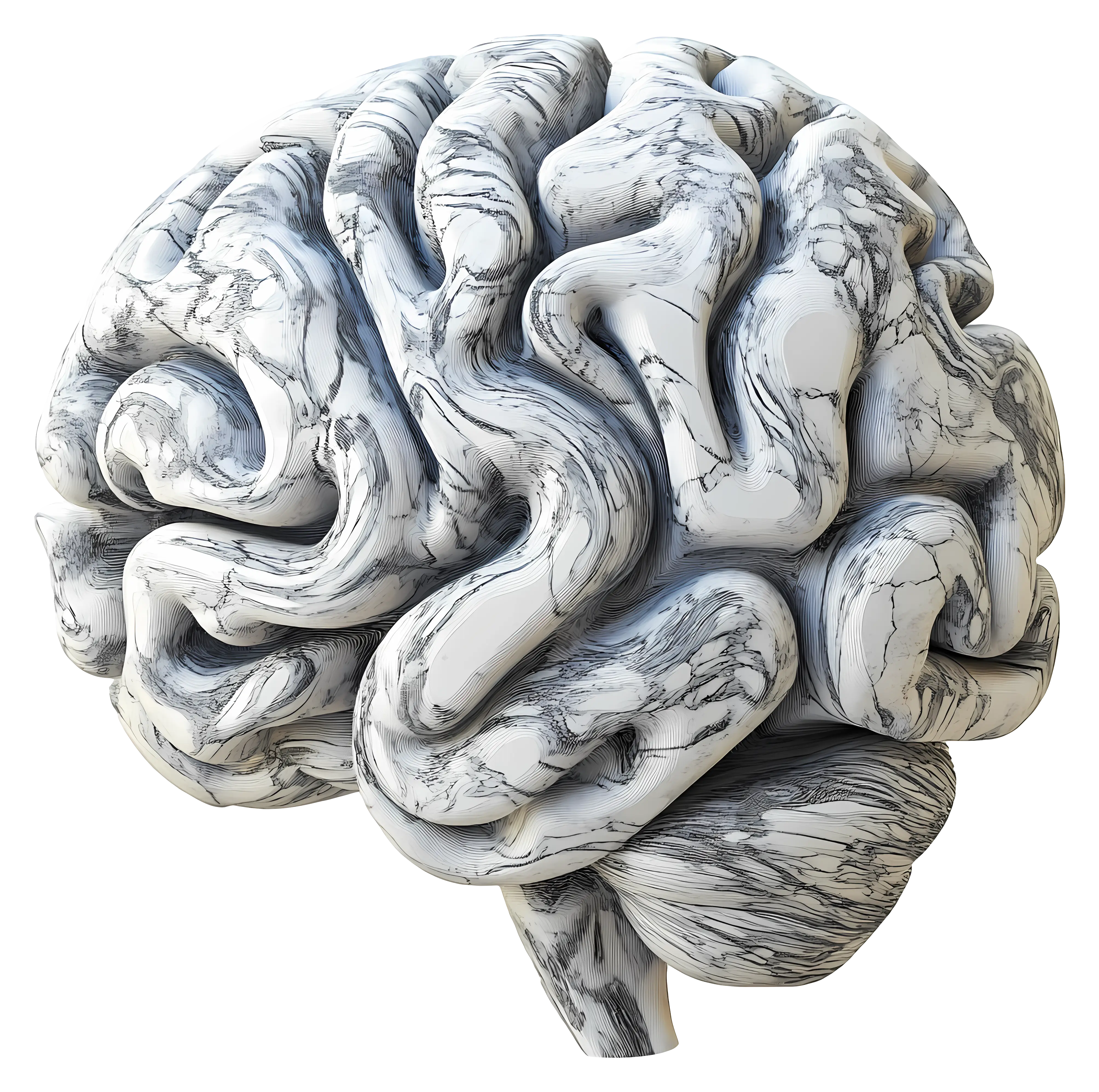
Marble Brain Illustration for Education -

Red Number Seven with Heart -

Gold Medal with Heart