You Might Like
-
Red Sharps Biohazard Container -
Chemical Molecule Structure Illustration -
Medical IV Catheter for Injection -
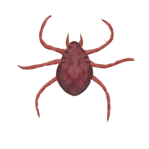
Brown Tick Illustration -

Cartoon Doctor with Stethoscope Illustration -
Yellow Skull and Bones Letter A Design -
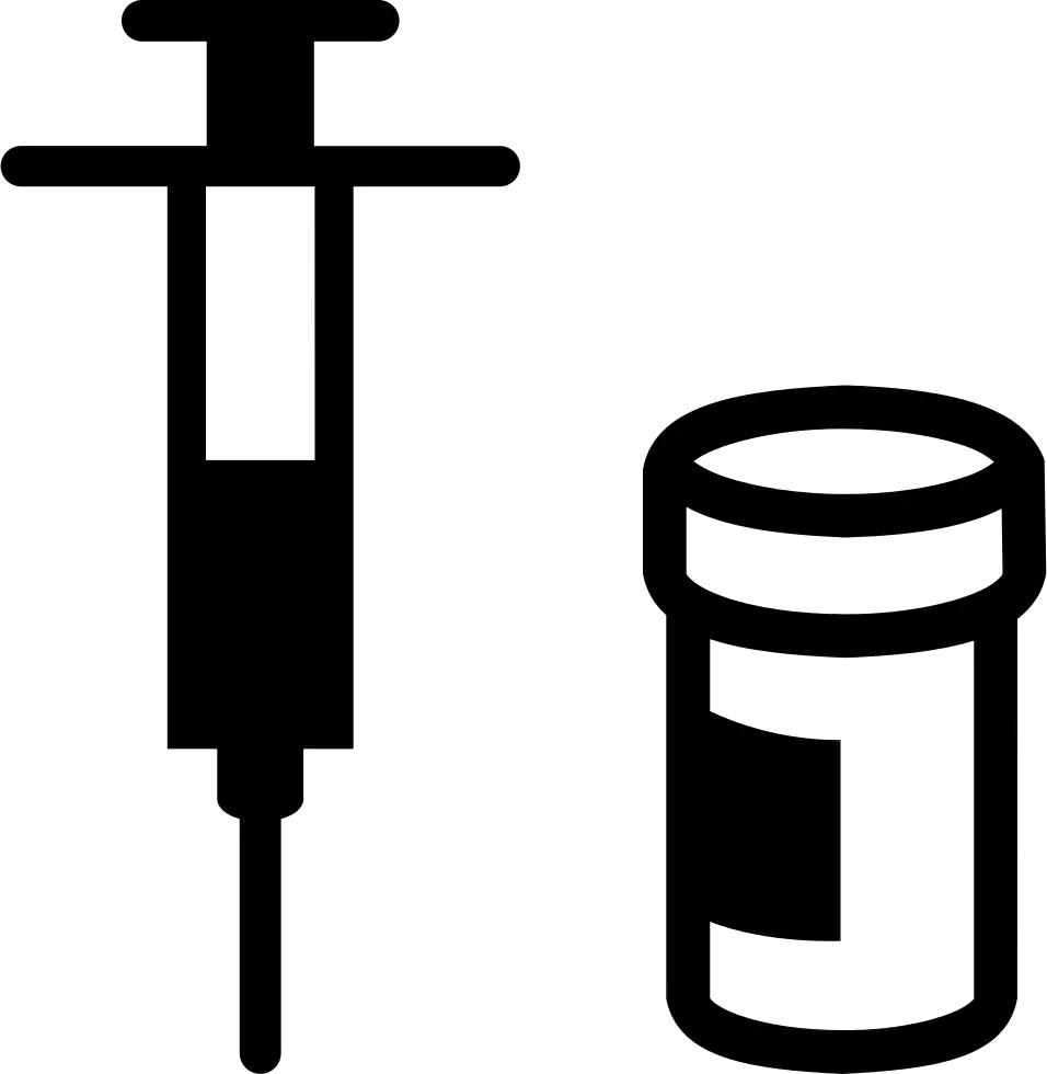
Syringe and Medicine Vial Icon -
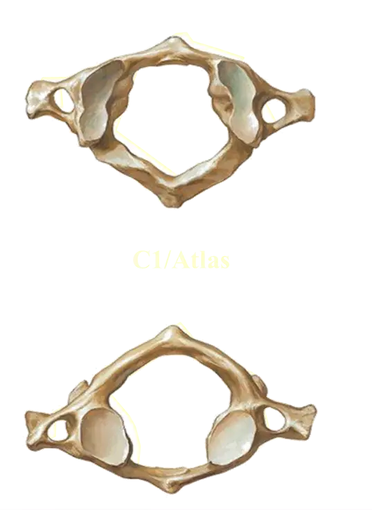
Atlas Vertebra Anatomy -

Dental Logo Design -
Abstract Colorful Skull Illustration -
Colorful Capsules and a Tablet Illustration -

Thermometer Symbol Icon -
Illustration of a Fish -

Strong Muscular Man Illustration -
Realistic Shark Illustration -
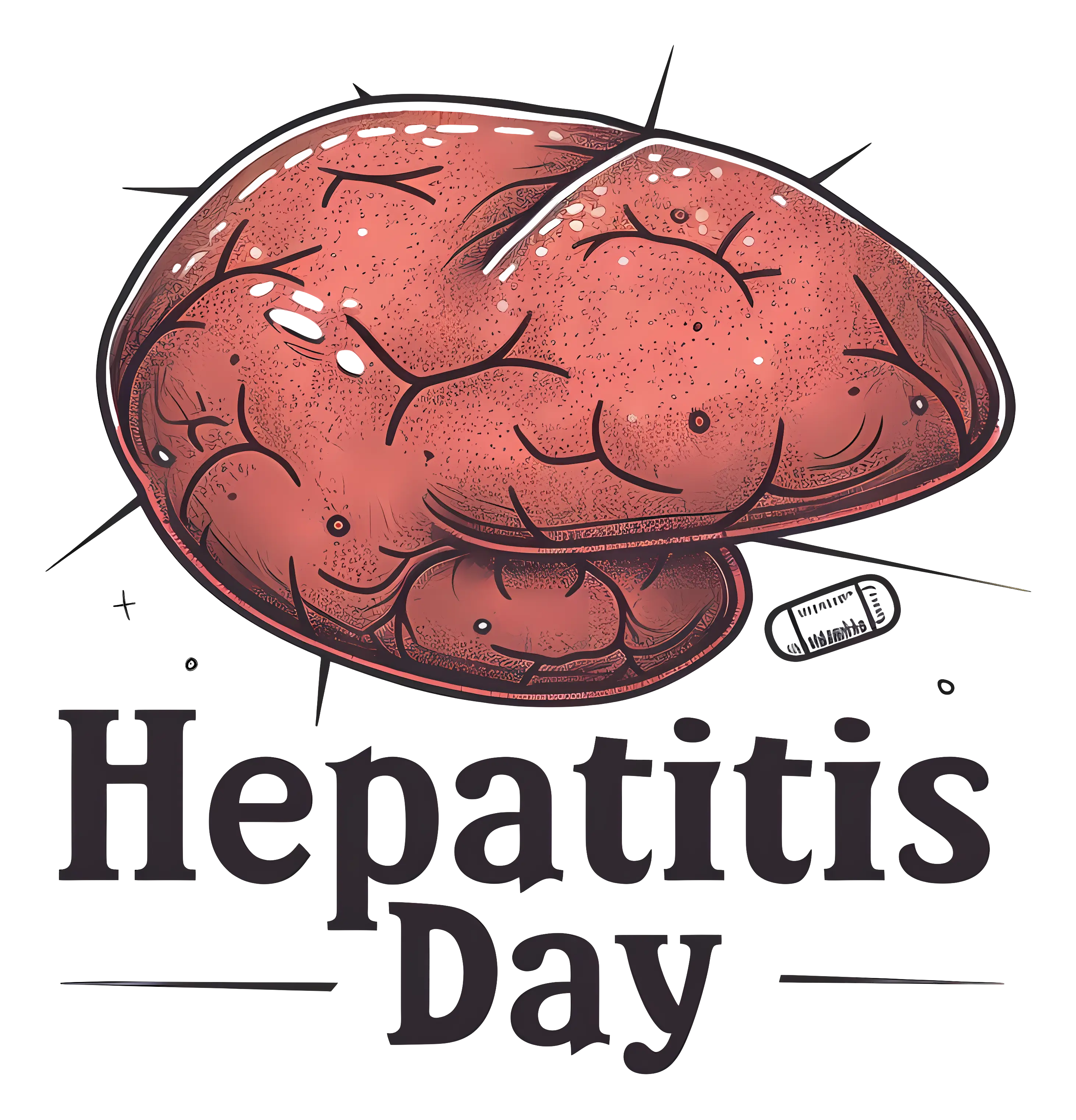
Hepatitis Day Awareness -
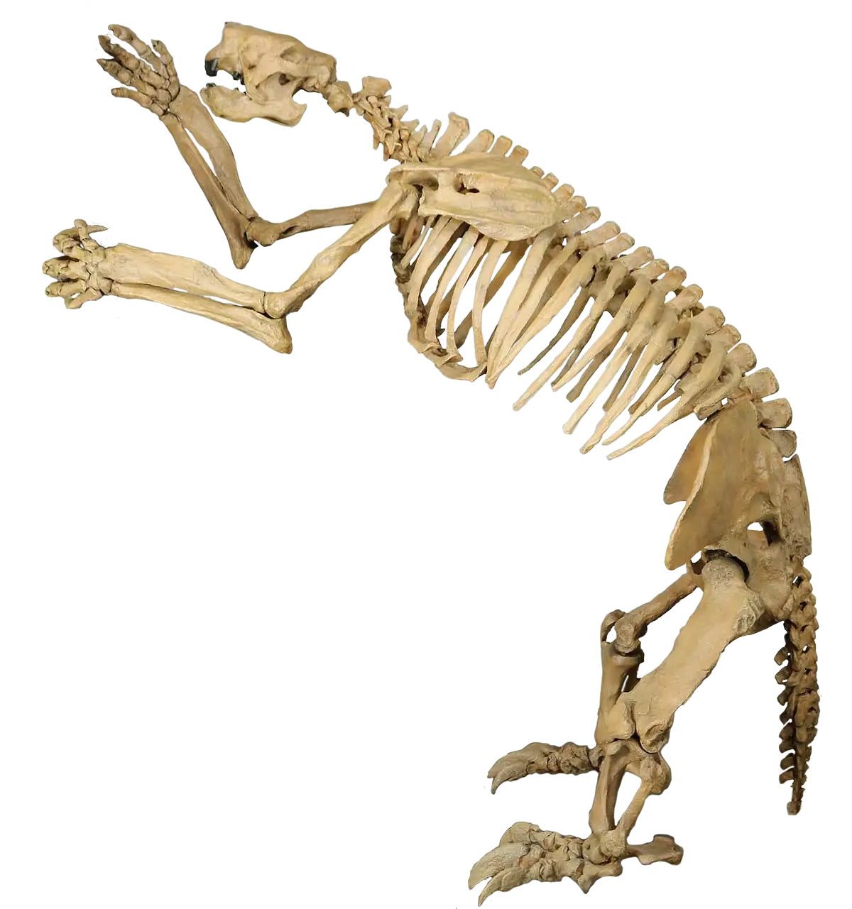
Animal Skeleton Display -

Medical Consultation Symbol -
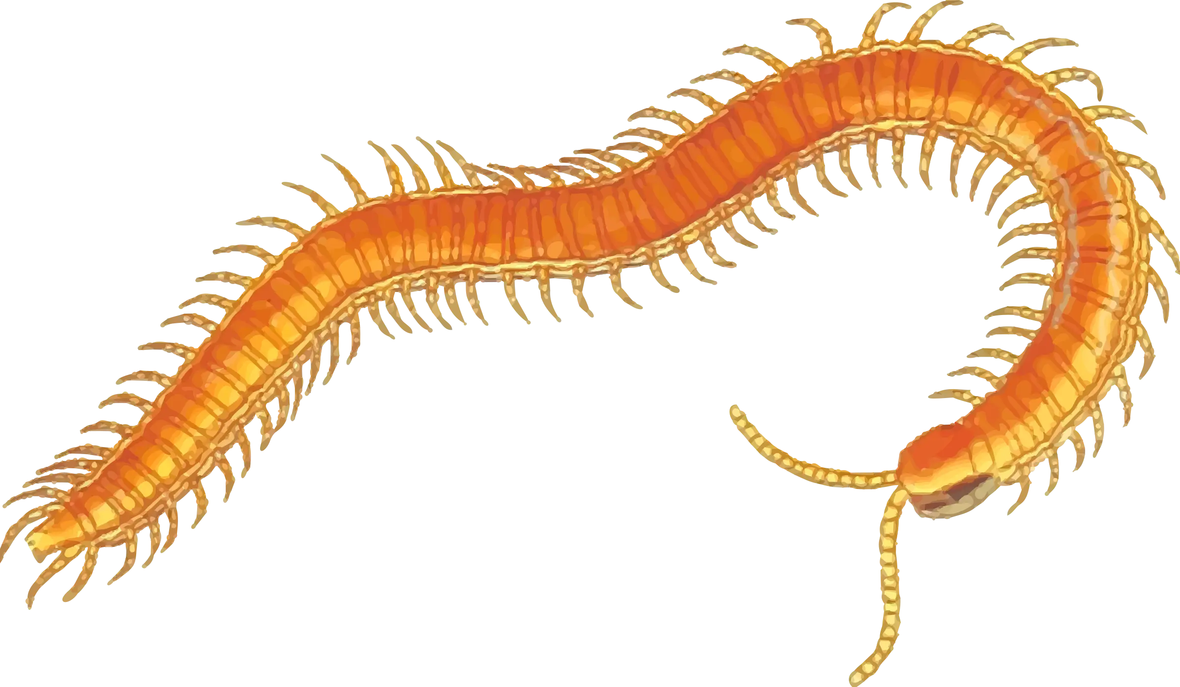
Orange Centipede Illustration -
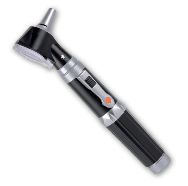
Medical Otoscope Instrument -

Medical Cross Icon in Red Background -
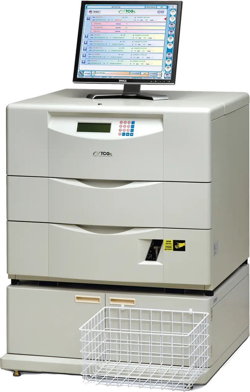
Advanced Medical Equipment -

Cartoon Doctors and Nurses Illustration -
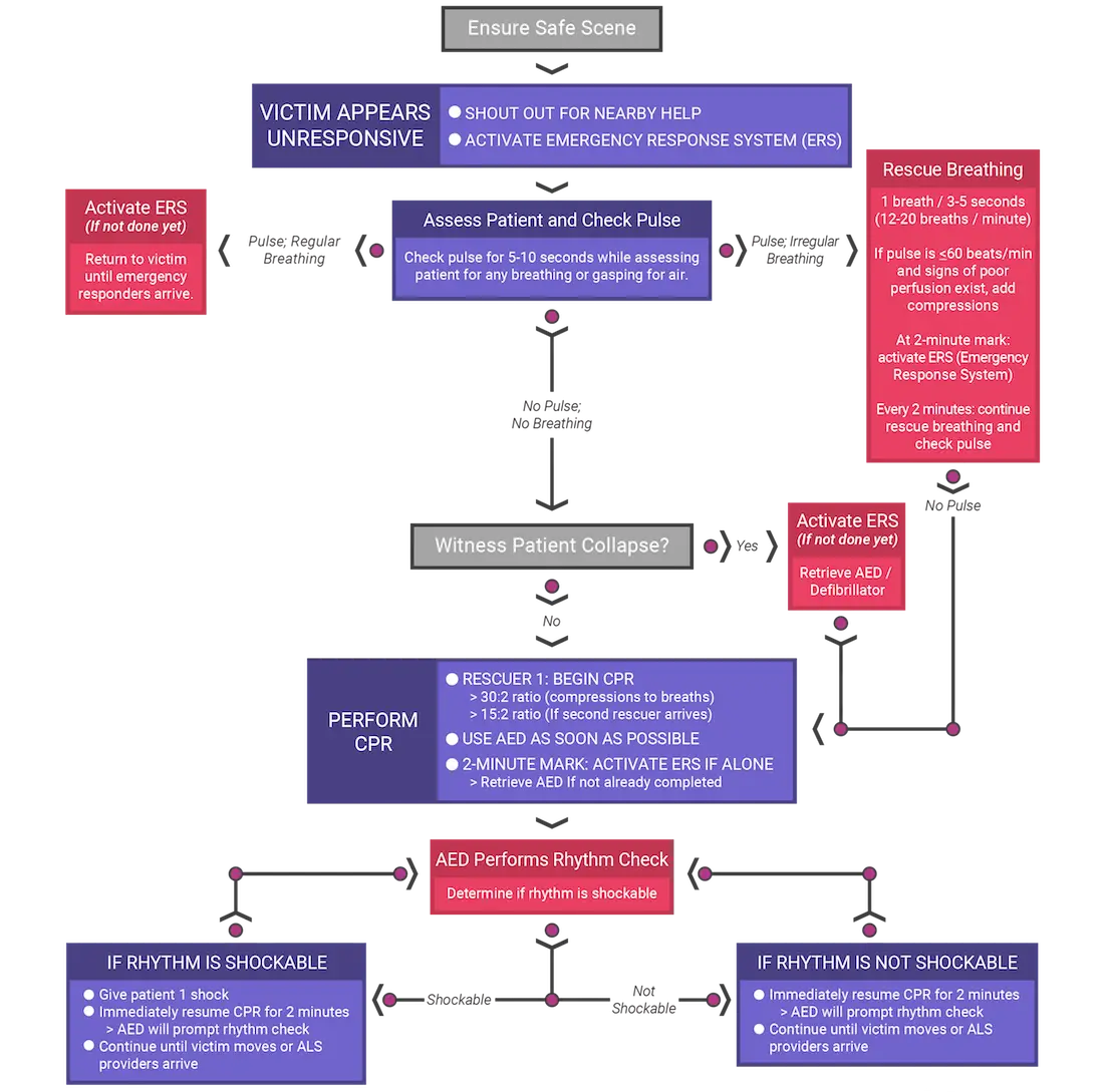
Medical CPR Flowchart Diagram -

Blue Human Brain Illustration -

Medical Cloud Symbol -
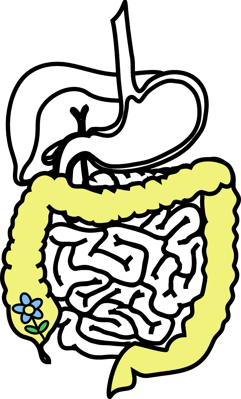
Digestive System Diagram -

Marble Brain Illustration for Education -

Silhouette of Black Hand in Vector Style -
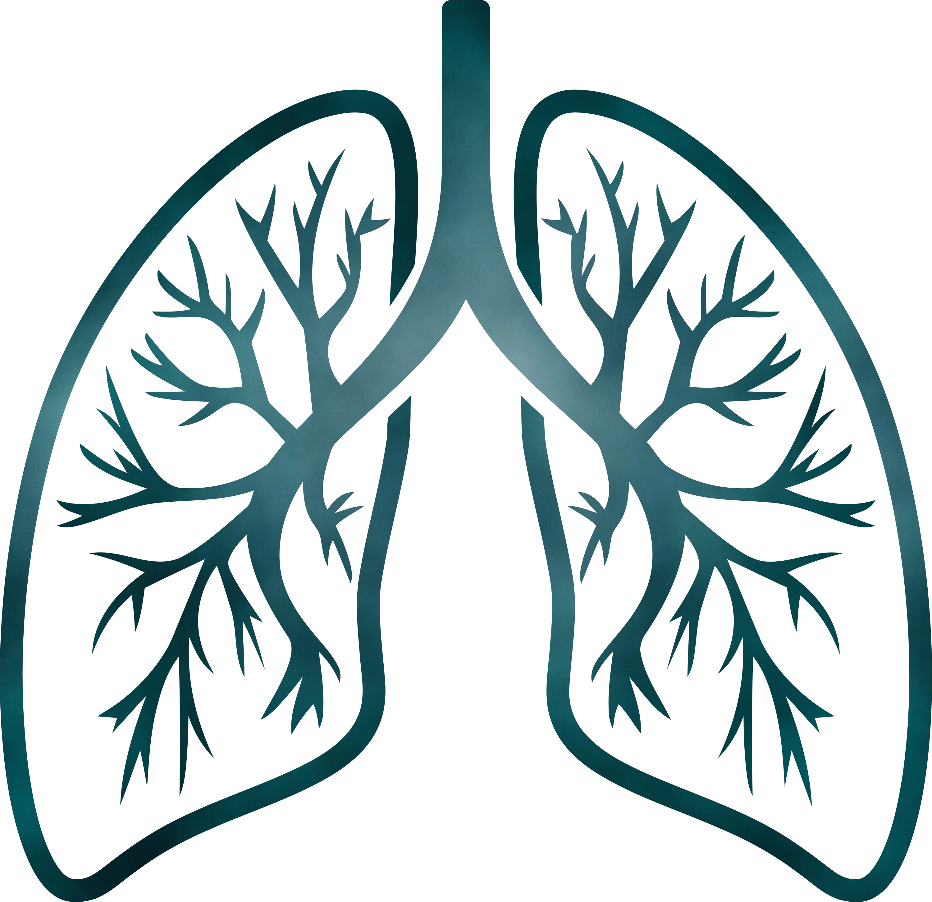
Detailed Lungs Diagram Illustration