You Might Like
-

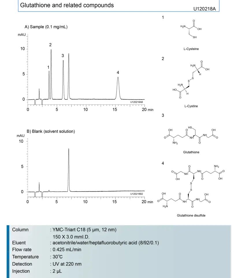
Scientific Chemical Structure Diagram -
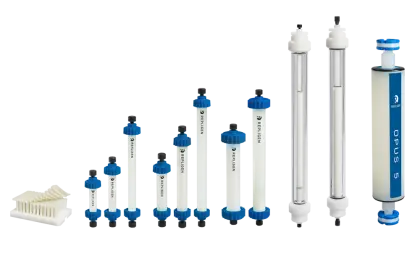
Laboratory Columns -
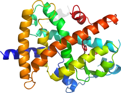
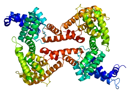
Protein Molecular Structure Model -
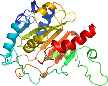
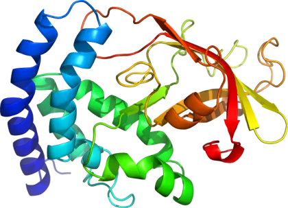
3D Protein Structure for Biochemistry Concepts -
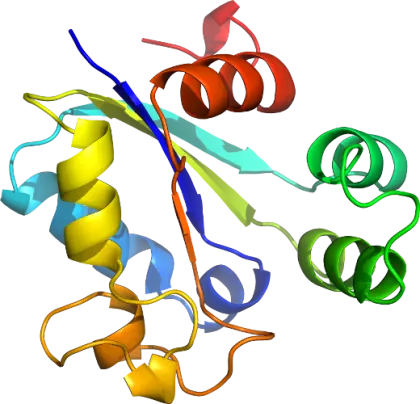
3D Protein Molecular Structure Illustration -
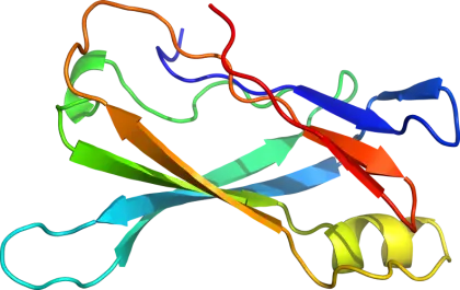
3D Protein Structure Illustration for Science -
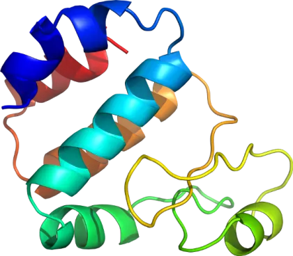
Colorful Protein Structure Illustration -
3D Representation of a Protein Structure -
3D Illustration of a Colorful Protein Structure -
3D Representation of Protein Structure -
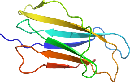
Colorful Protein Structure Illustration -
Protein Structure Diagram in Molecular Biology -
Protein Structure Scientific Illustration -
Colorful Protein Structure Illustration -
Colorful Protein Structure Illustration for Science -
3D Ribbon Structure of a Protein Molecule -
Protein Complex 3D Representation -
Scientific Illustration of Protein Structure -
3D Protein Structure Molecule Diagram -
Colorful Representation of Protein Structure -
Molecular Protein Structure Illustration -
Colorful Protein Structure Illustration