You Might Like
-
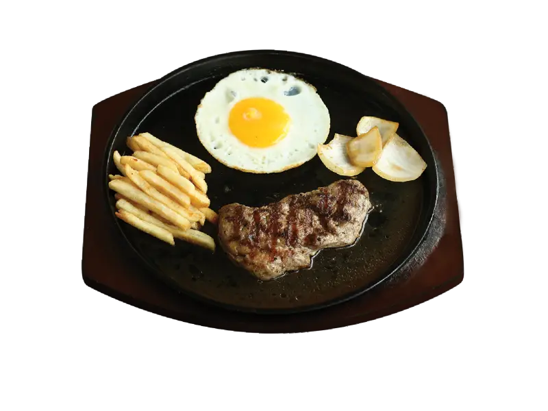
Steak and Egg Meal with Fries -
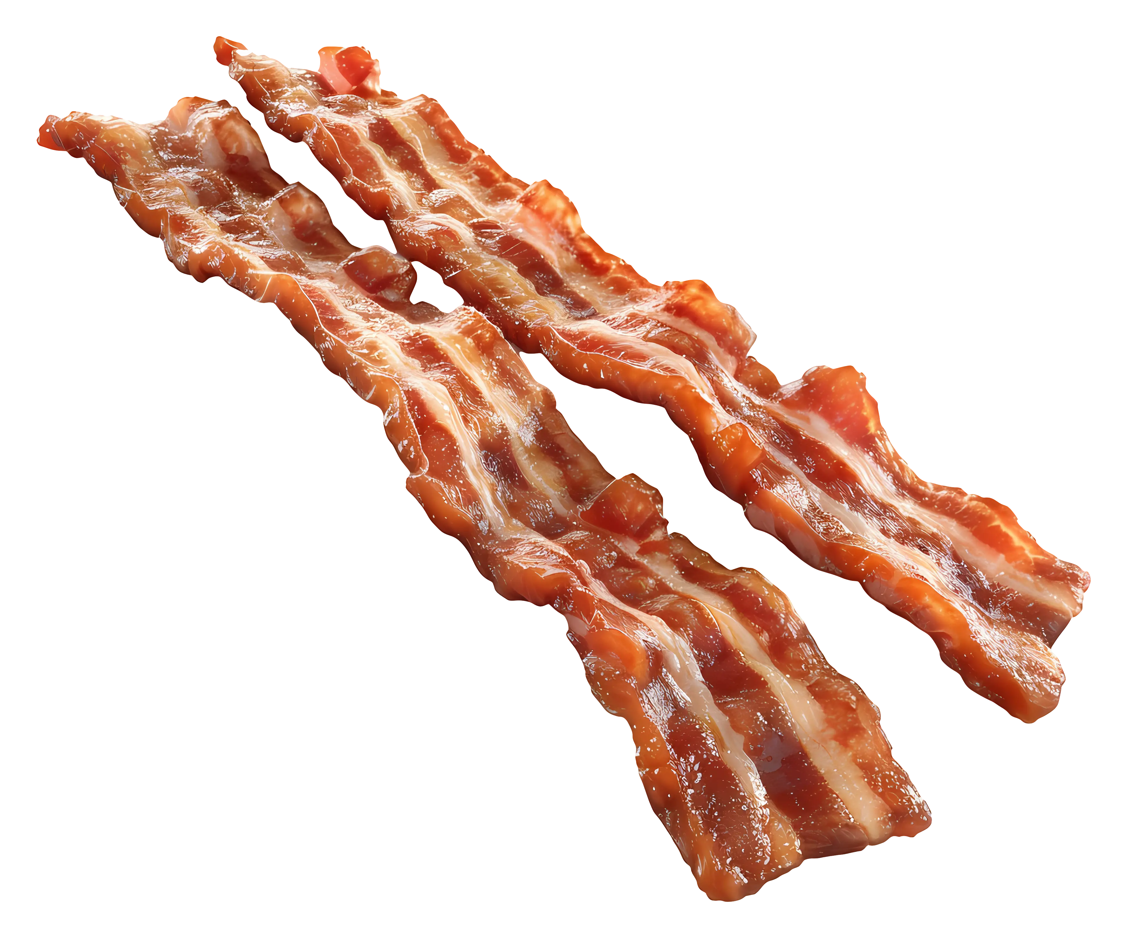
Illustration of Crispy Bacon Strips -
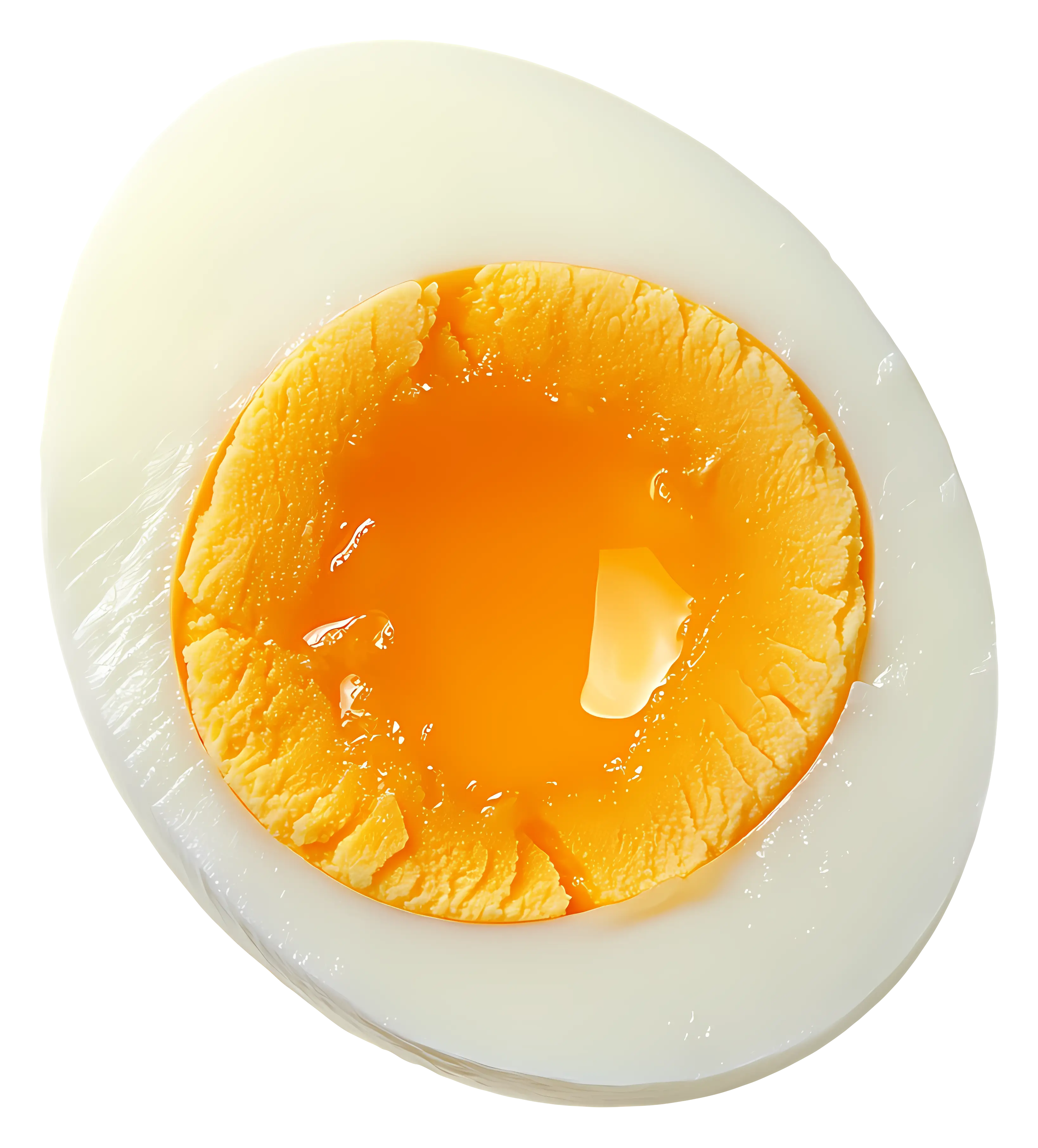
Half Boiled Egg with Soft Yolk -
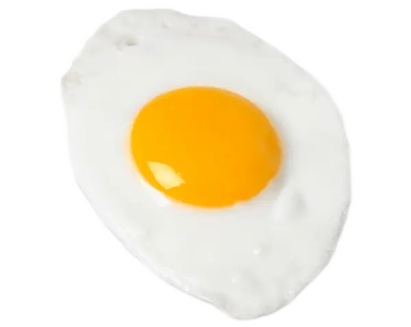
Fried Egg Illustration -
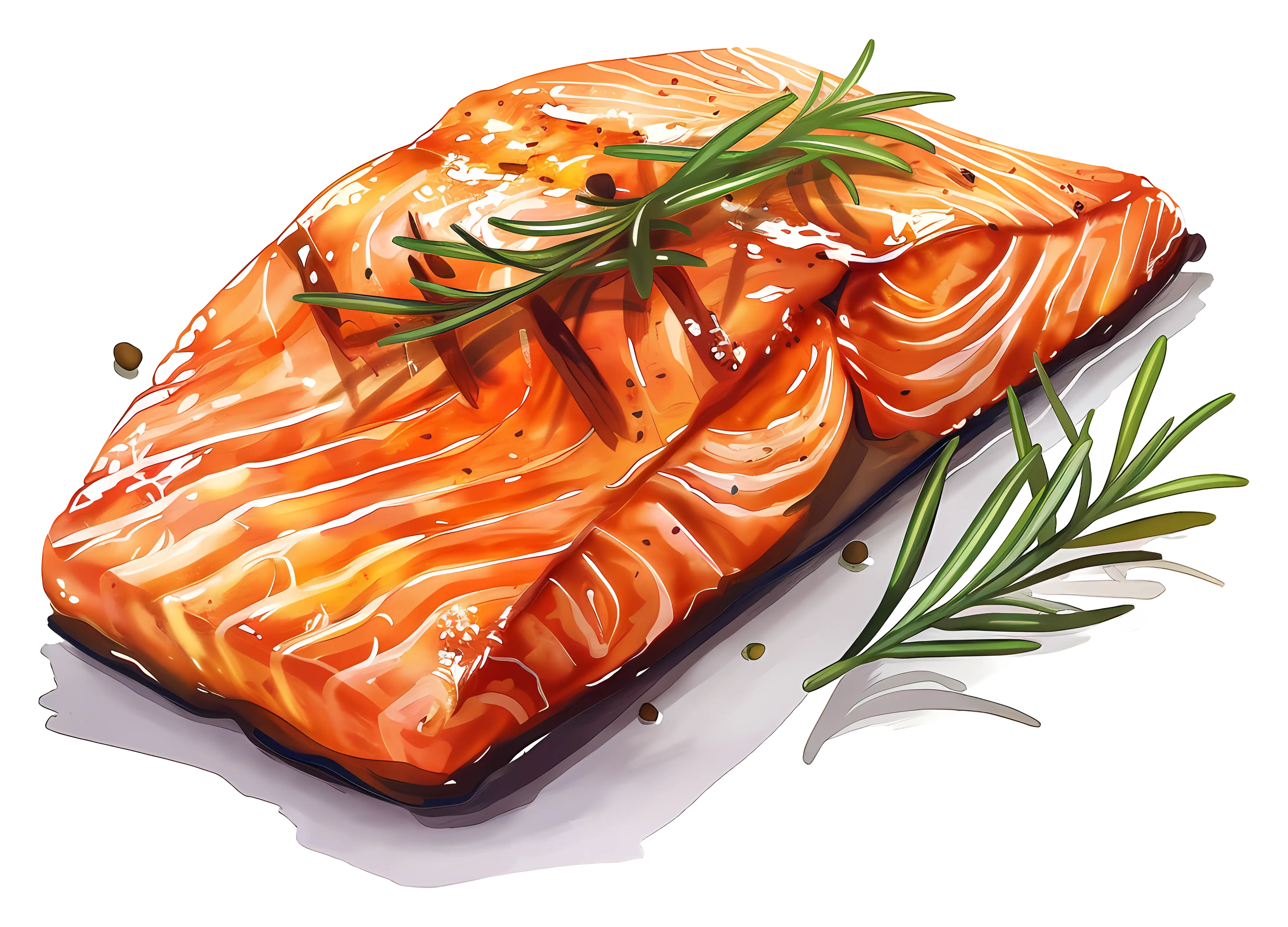
Roasted Salmon with Rosemary Garnish -

Fried Egg with Garnish Illustration -
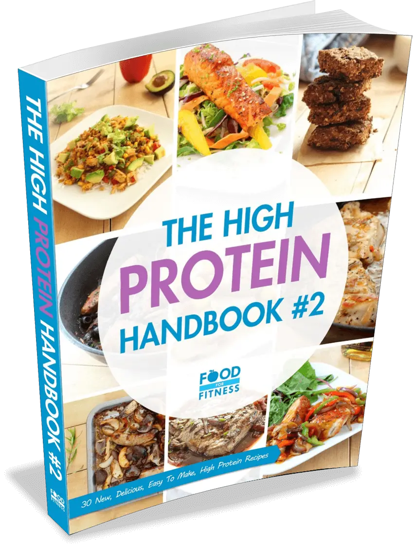
The High Protein Handbook #2 -
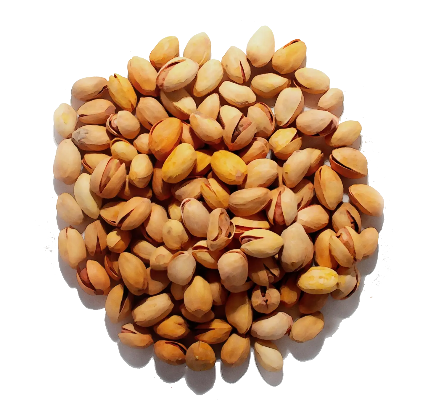
Pile of Pistachio Nuts -

Scientific Chemical Structure Diagram -
Chicken Patties with Herbs -
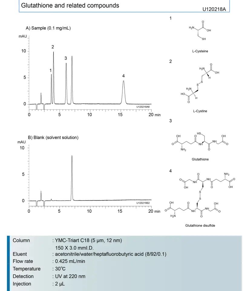
Chemical Analysis and Data Representation -
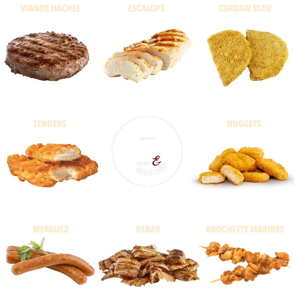
Assorted Meat Food Items -
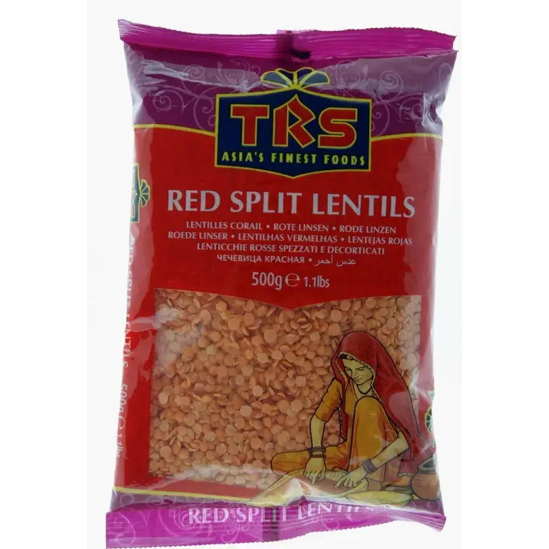
TRS Red Split Lentils Package -
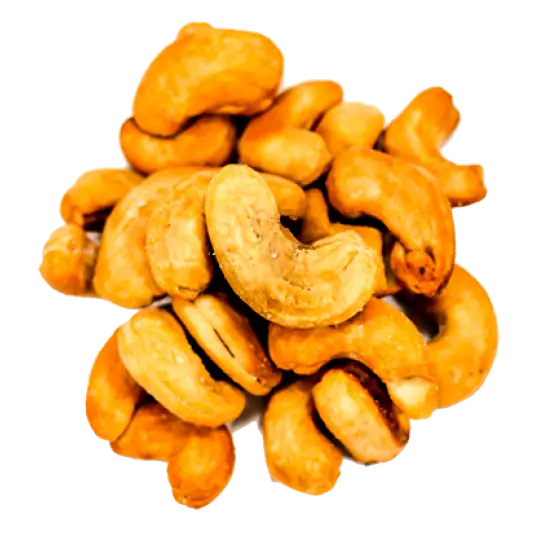
Crunchy Roasted Cashew Nuts for Healthy Snacking -
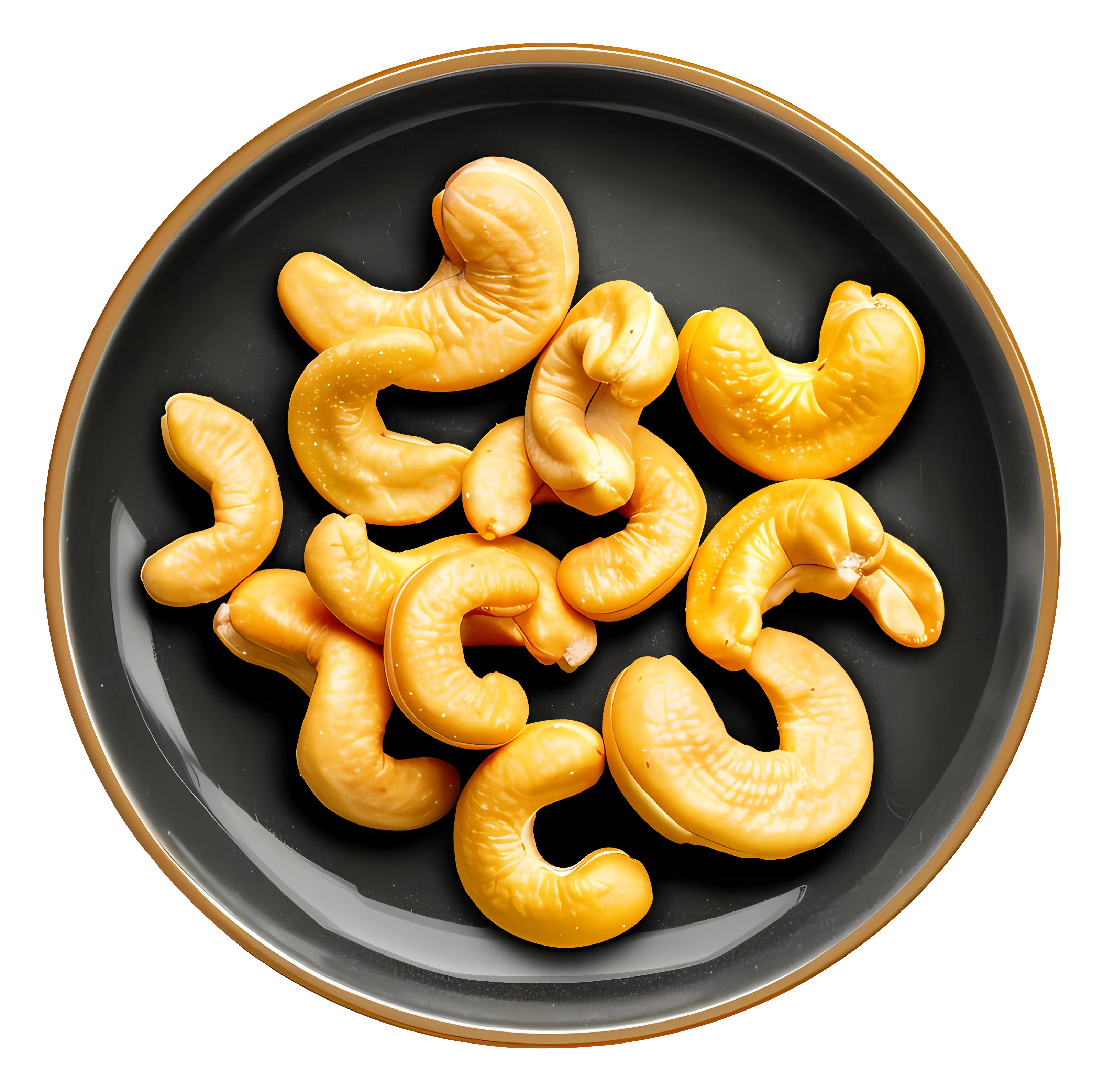
Cashew Nuts Served in a Black Bowl -
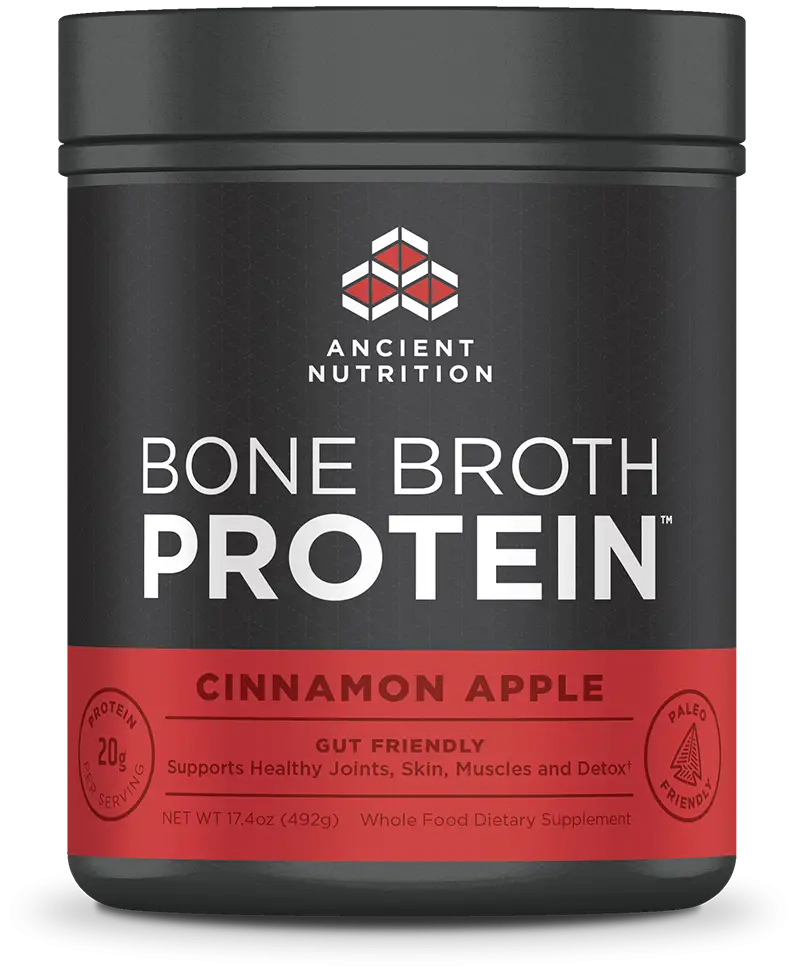
Bone Broth Protein Jar in Cinnamon Apple Flavor -
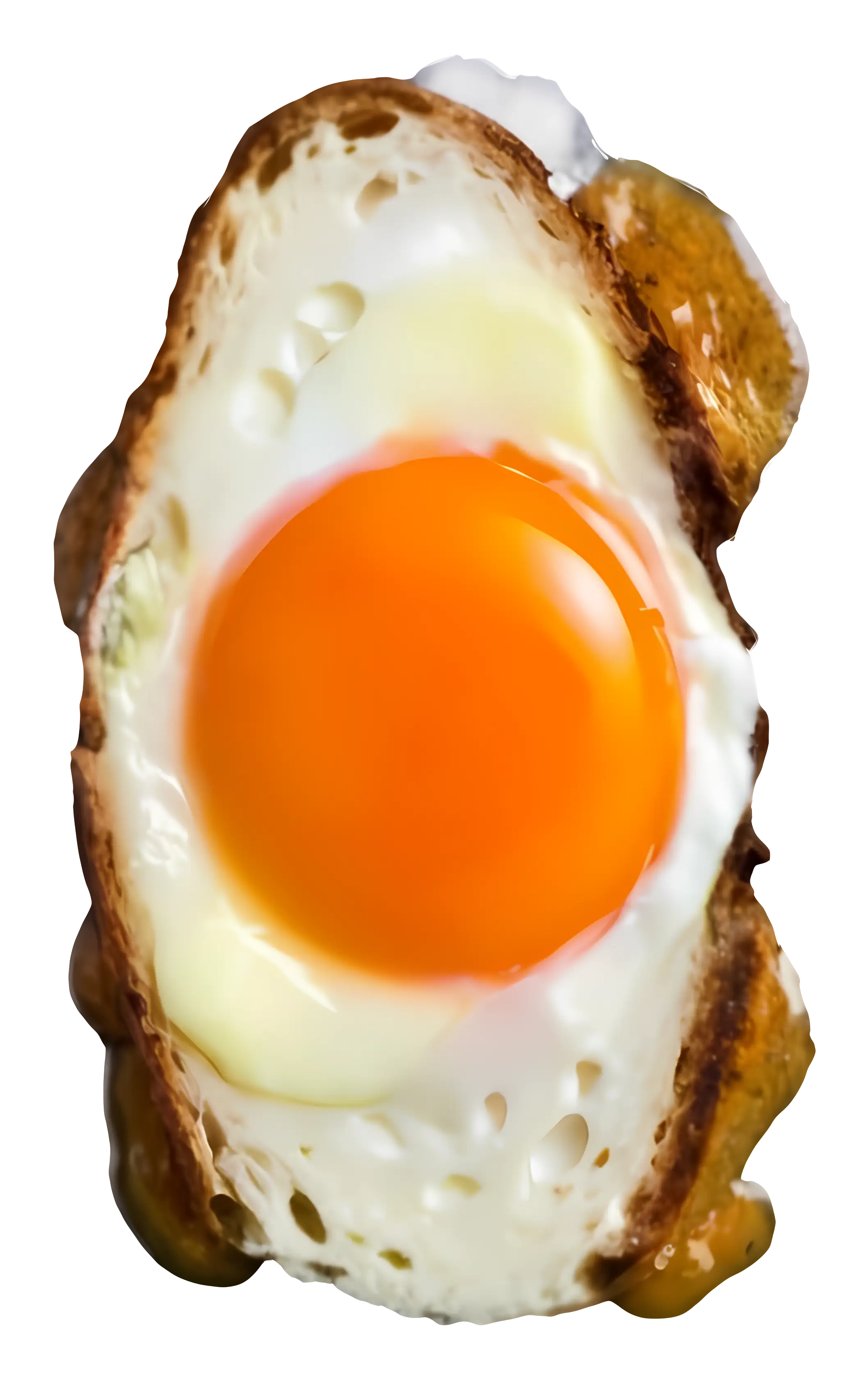
Realistic Fried Egg -
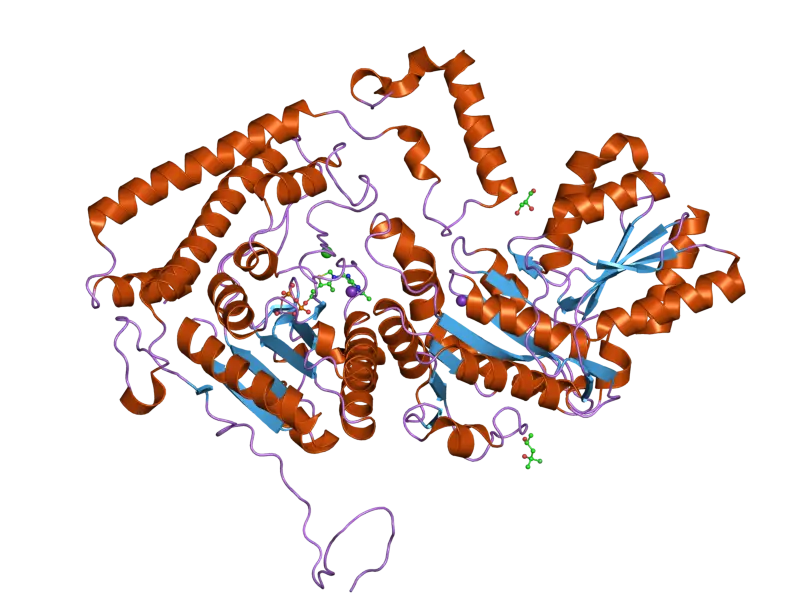
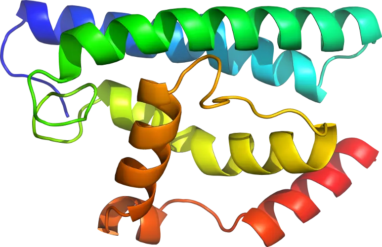
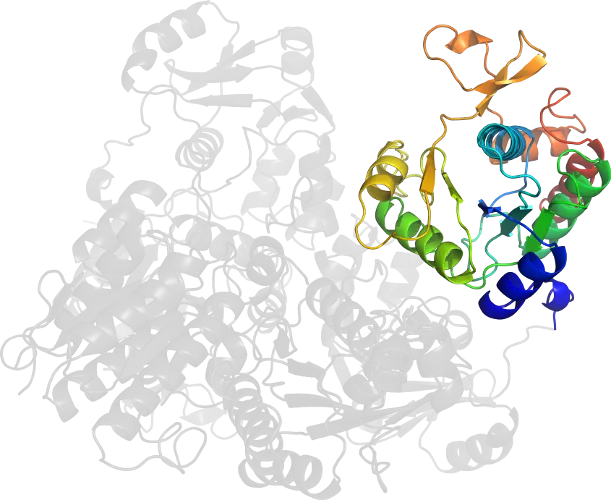
Protein Molecular Structure Model -

Boiled Egg with Soft Yolk -
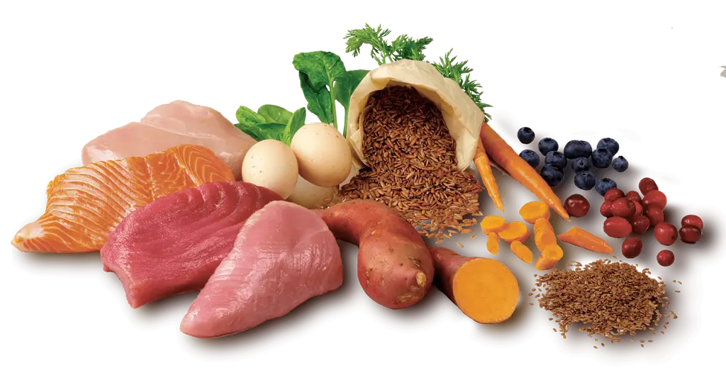
Assorted Fresh Food Ingredients -
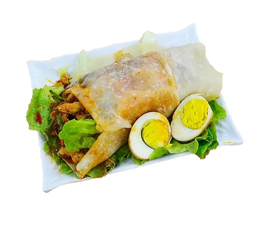
Delicious Food Dish with Eggs and Salad -
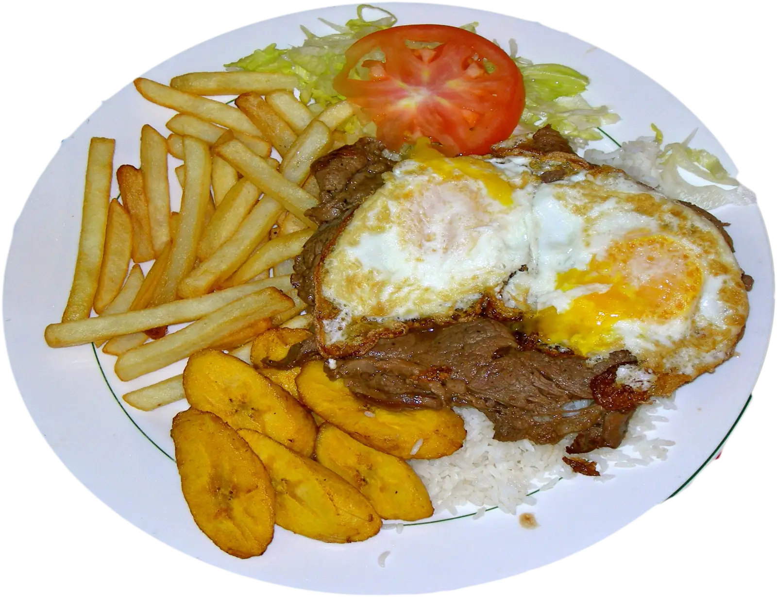
Delicious Meal with Fries and Eggs -
Casein Protein Powder Container for Fitness Enthusiasts -
Packaged Turkey and Bacon Salad -
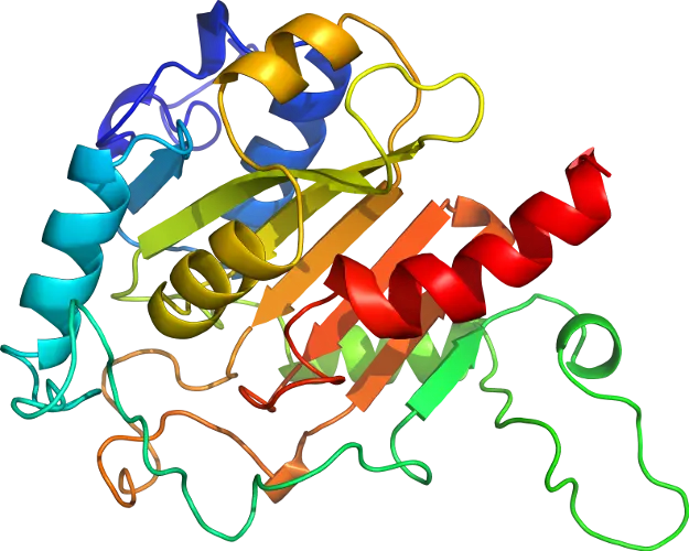
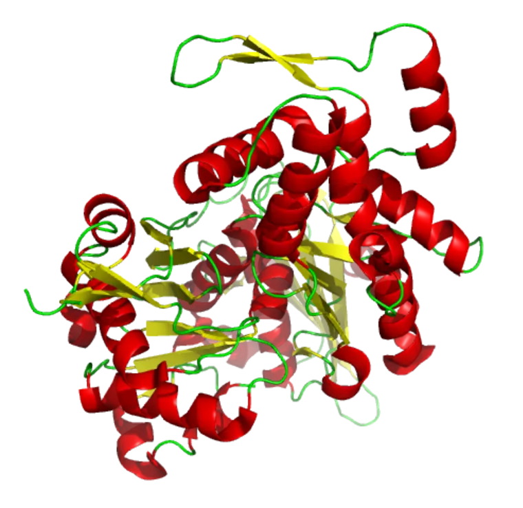
3D Protein Structure for Biochemistry Concepts -

3D Protein Molecular Structure Illustration -
3D Protein Structure Illustration for Science -
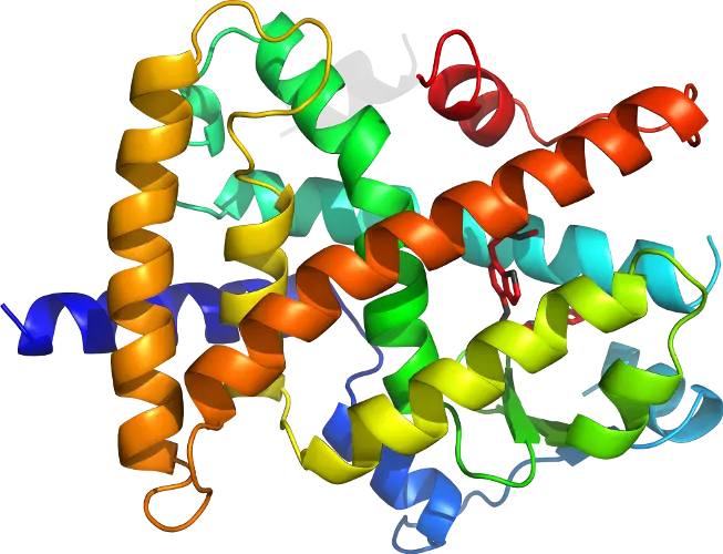
Colorful Protein Structure Illustration -

Pile of Raw Soybeans -
3D Representation of a Protein Structure