You Might Like
-
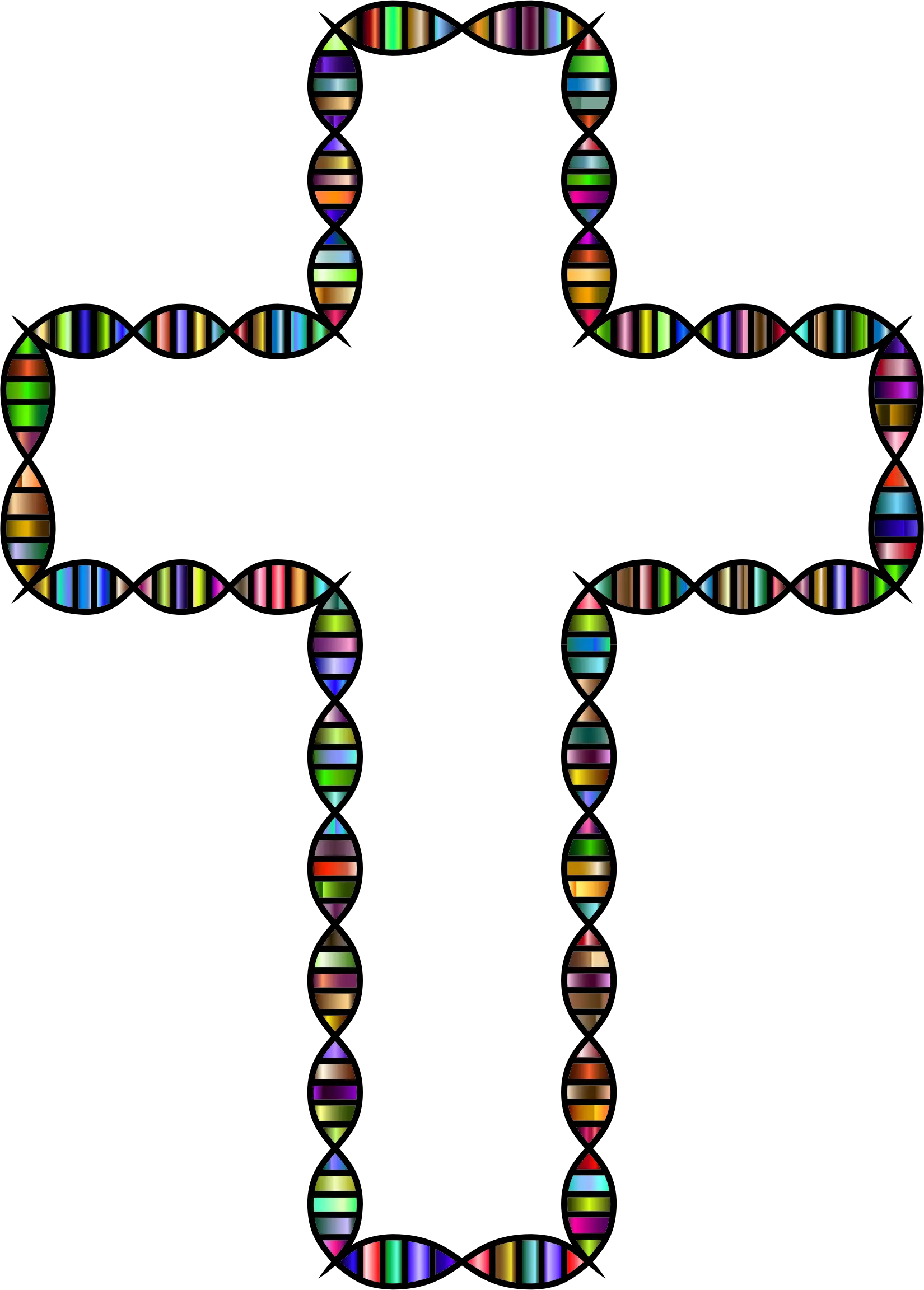
Cross Made of DNA Strands -

Hand-drawn DNA Stranded Illustration -

Colorful DNA Strand Illustration -
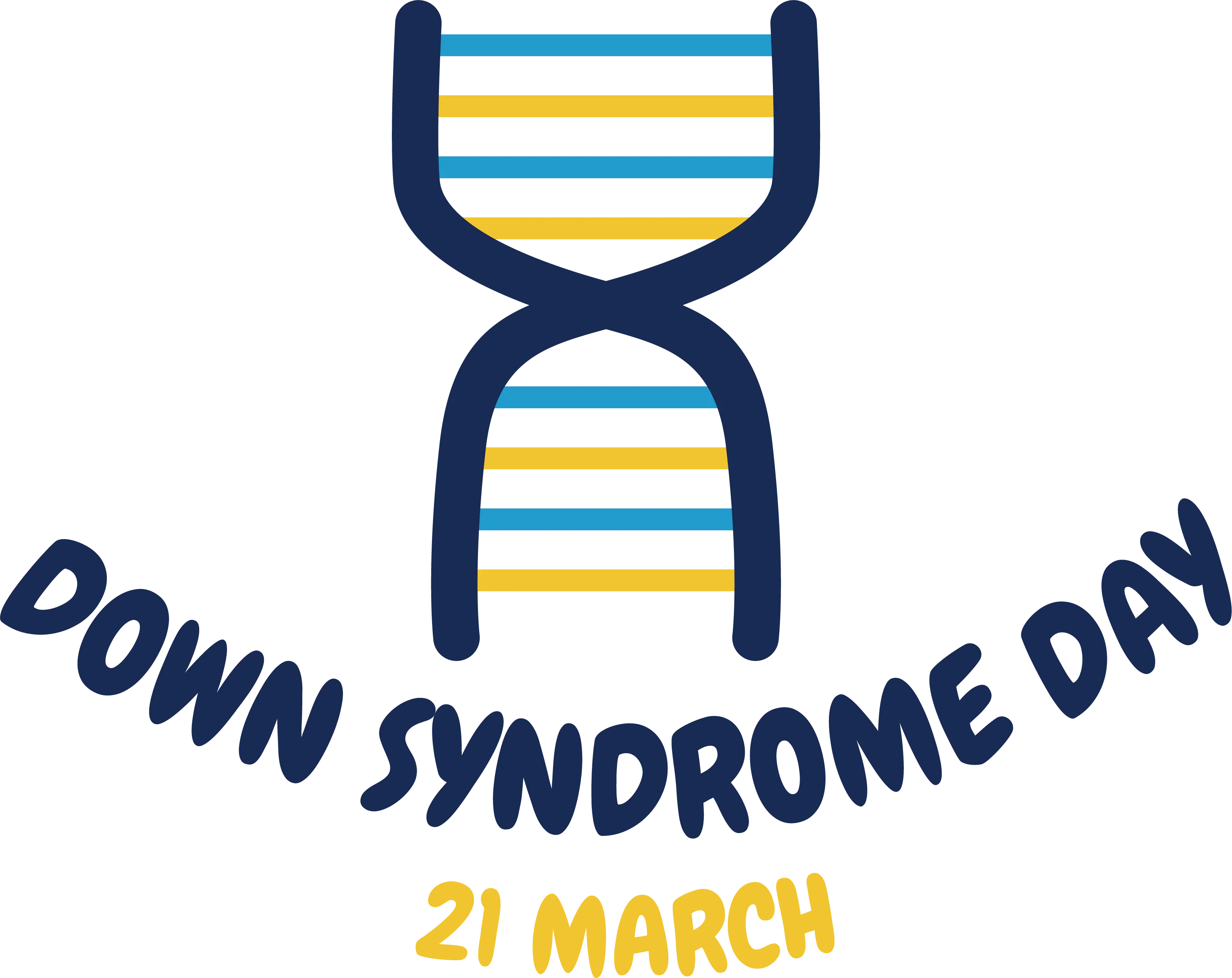
DNA Symbol for Down Syndrome Day -
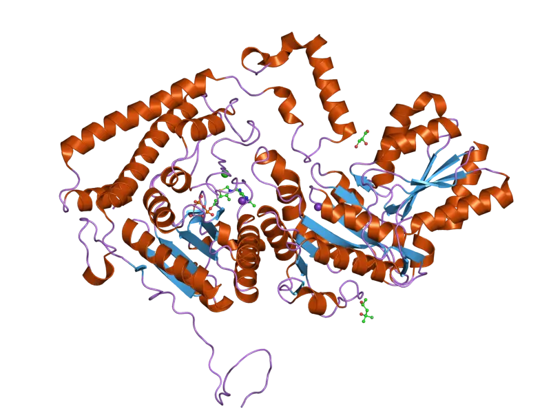
Protein Structure Diagram in Molecular Biology -
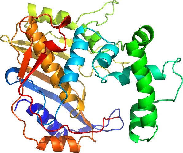
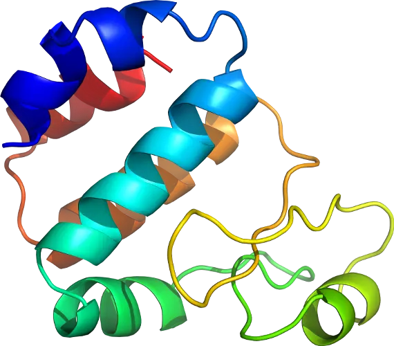
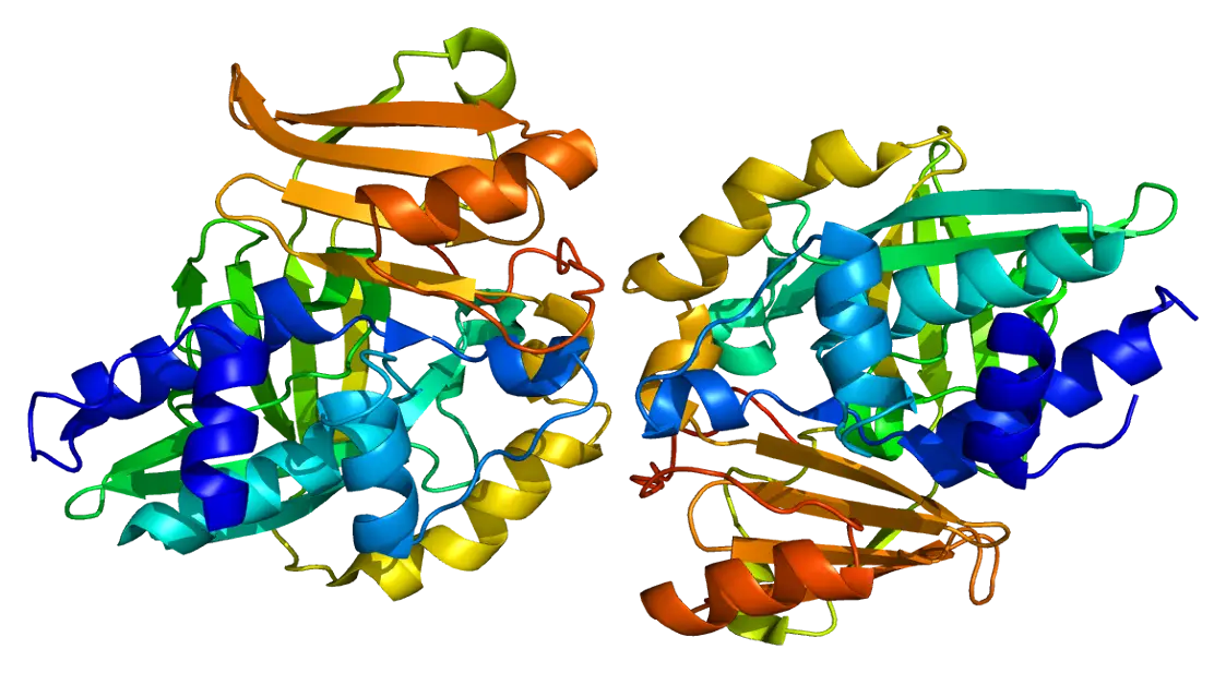
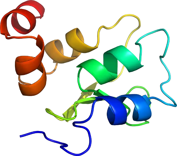
Colorful Protein Structure Illustration -
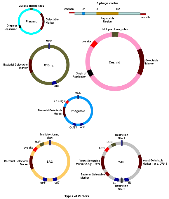
Types of Vectors in Genetic Engineering -

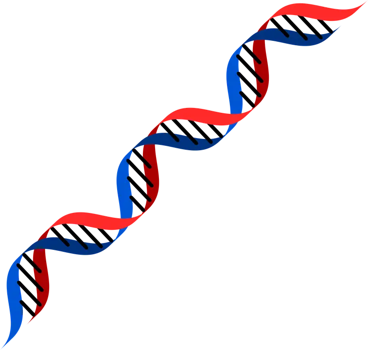
Double Helix DNA Strand Illustration -
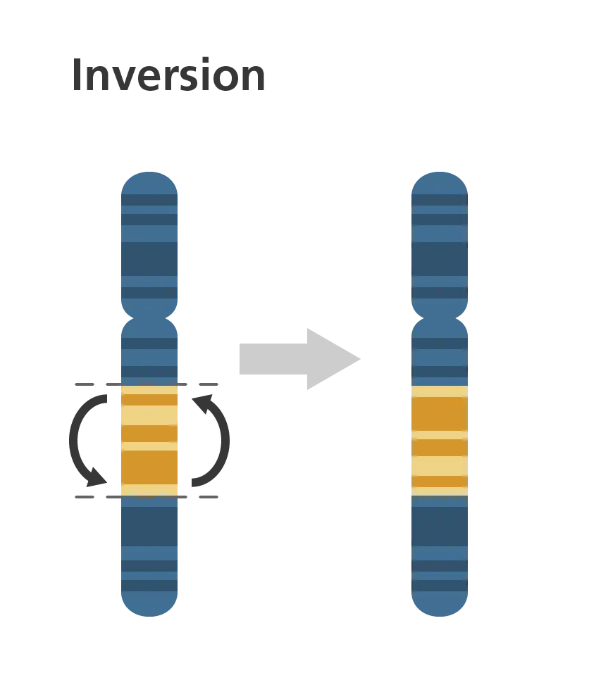
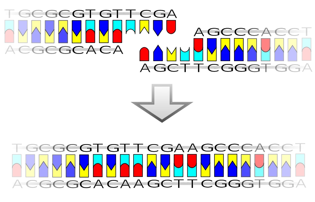
Chromosome Inversion Diagram -
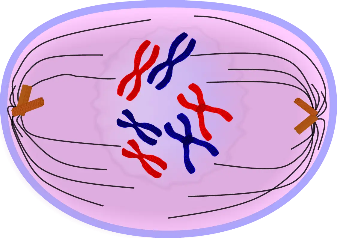
Chromosomes in Cell Division Illustration -
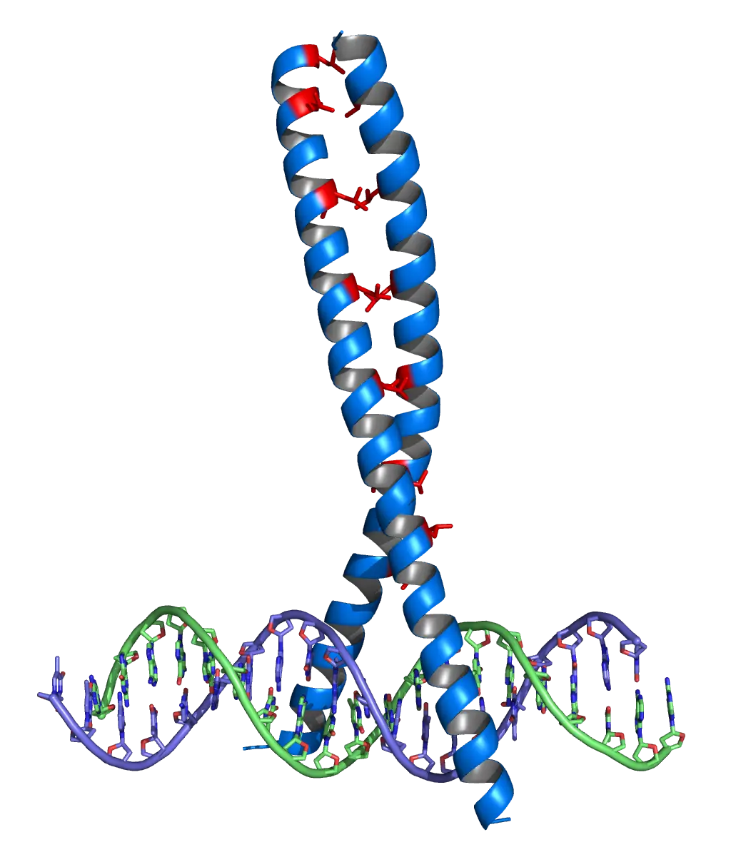
DNA and Protein Structure Illustration -
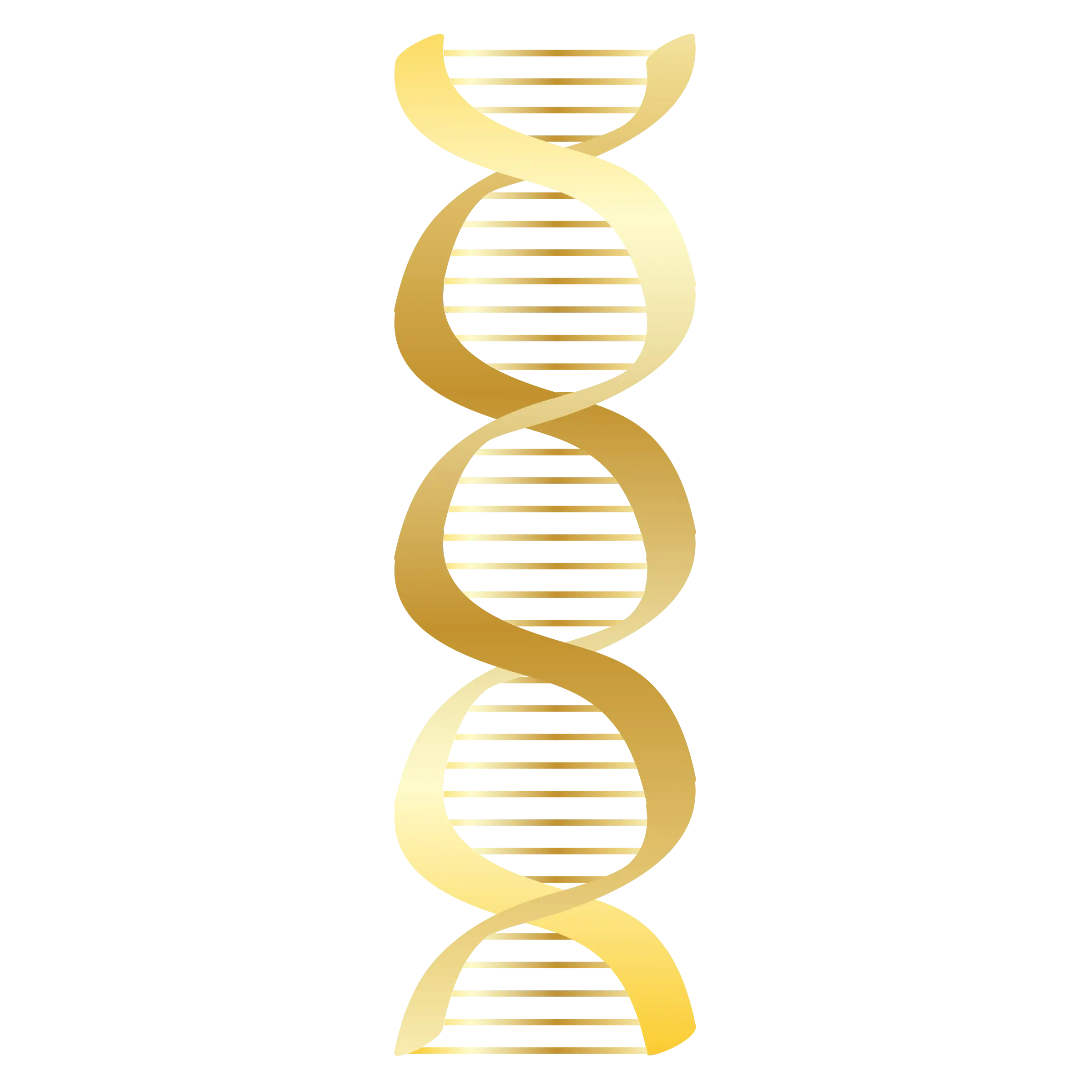
Golden DNA Helix Model for Science and Decoration -
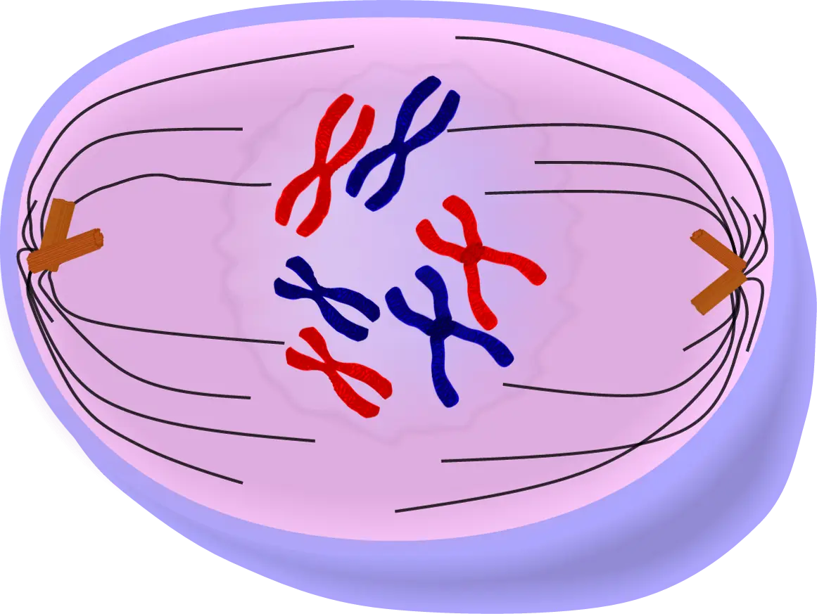
Illustration of Cell Division Showing Chromosomes -
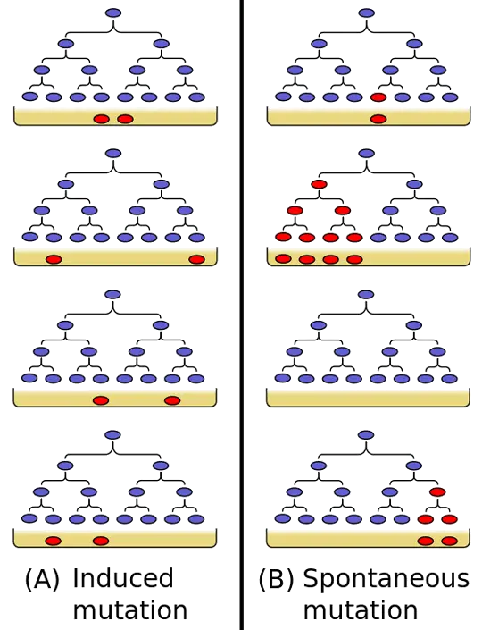
Diagram of Induced and Spontaneous Mutations -
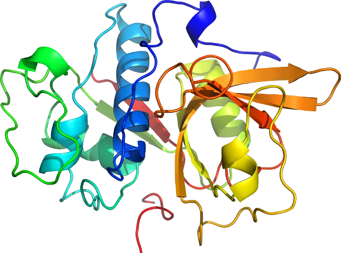
Molecular Protein Structure Illustration -
Red DNA Helix Symbol -

Blue Double-Helix DNA Strand Illustration -
Protein Ribbon Structure for Molecular Biology Visualization -

Blue DNA Spiral and Green Leaf Logo -
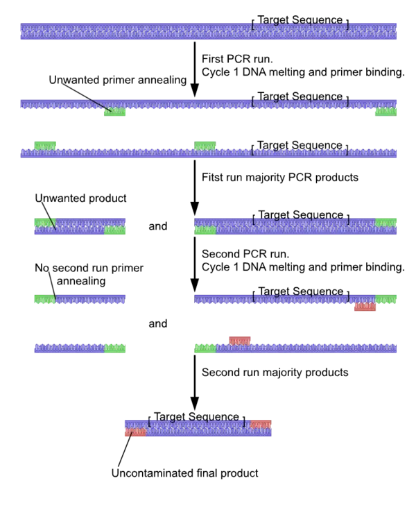
PCR Process Diagram for Science Education -

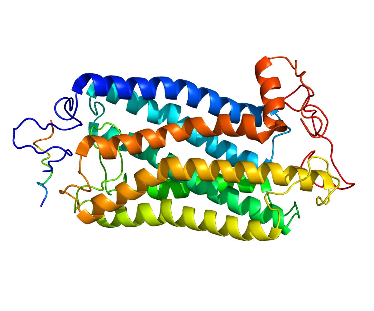
Colorful Protein Structure Model -
DNA Double Helix Illustration -
Protein Structure Scientific Diagram -
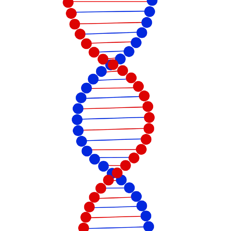
Colorful DNA Double Helix Illustration -
Colorful Protein Molecular Structure -
Protein 3D Structure in Biochemistry -
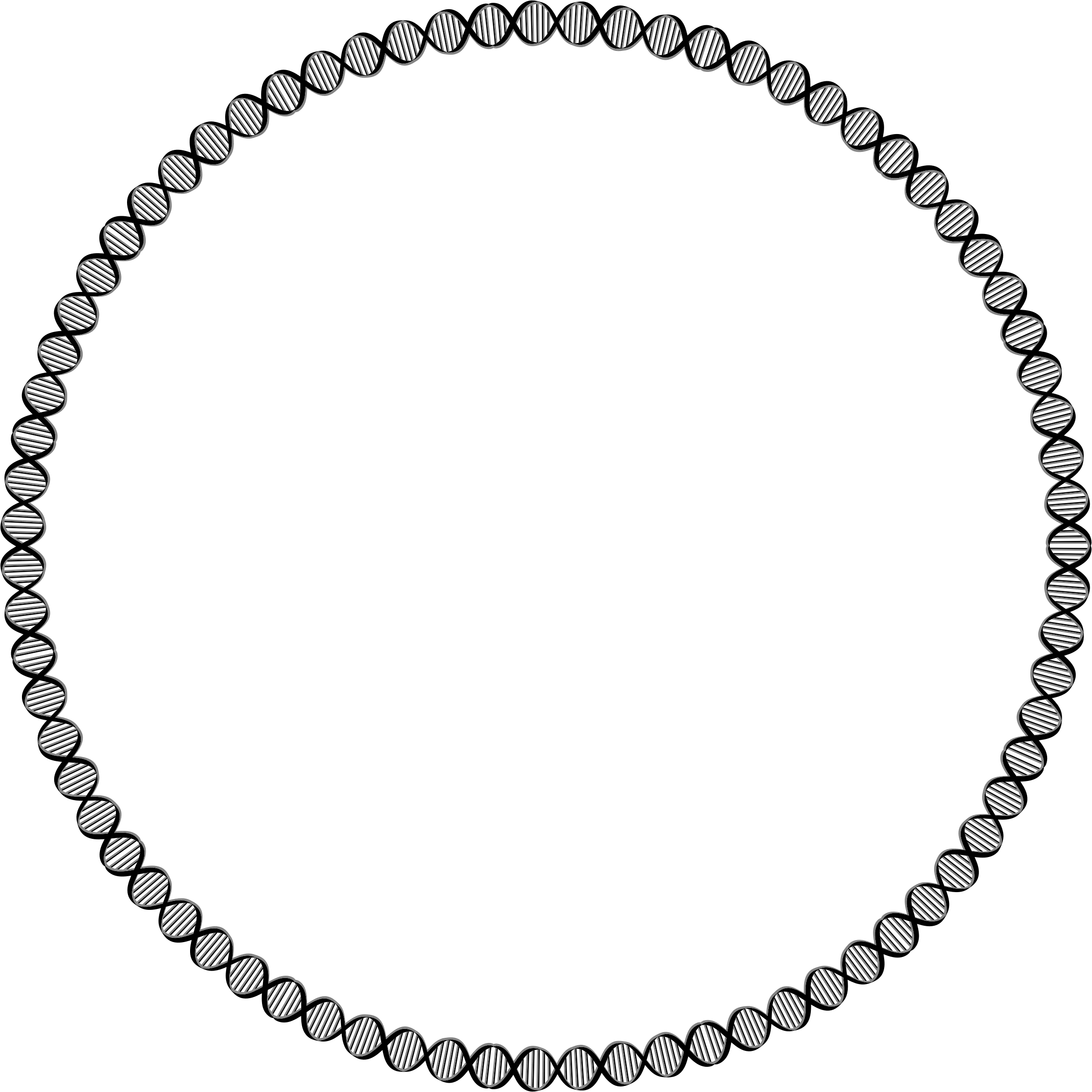
Decorative Circular DNA Frame -

2'-Deoxycytidine Molecule Structure -
3D Representation of a Protein Structure -
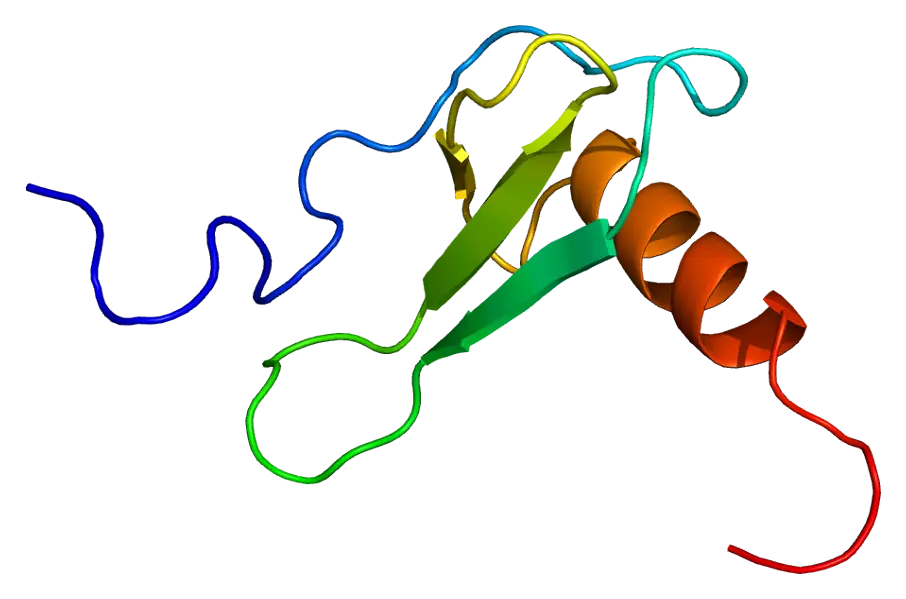
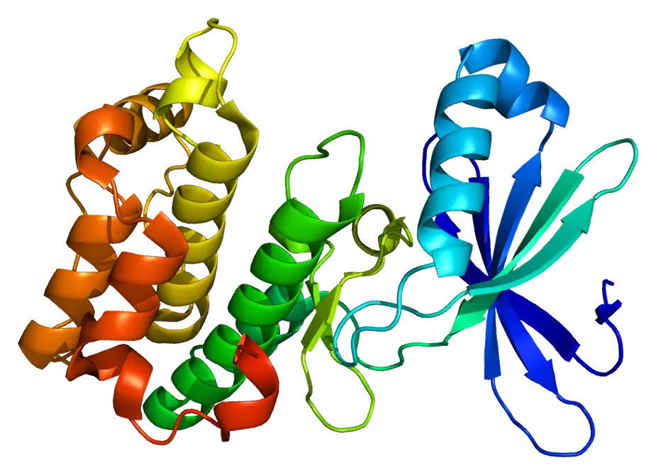
Protein Ribbon Structure Representation