You Might Like
-
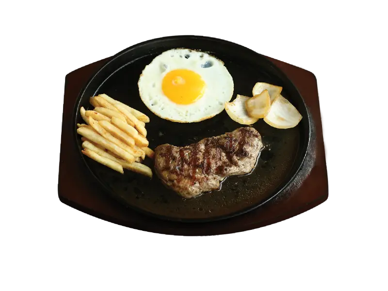
Steak and Egg Meal with Fries -
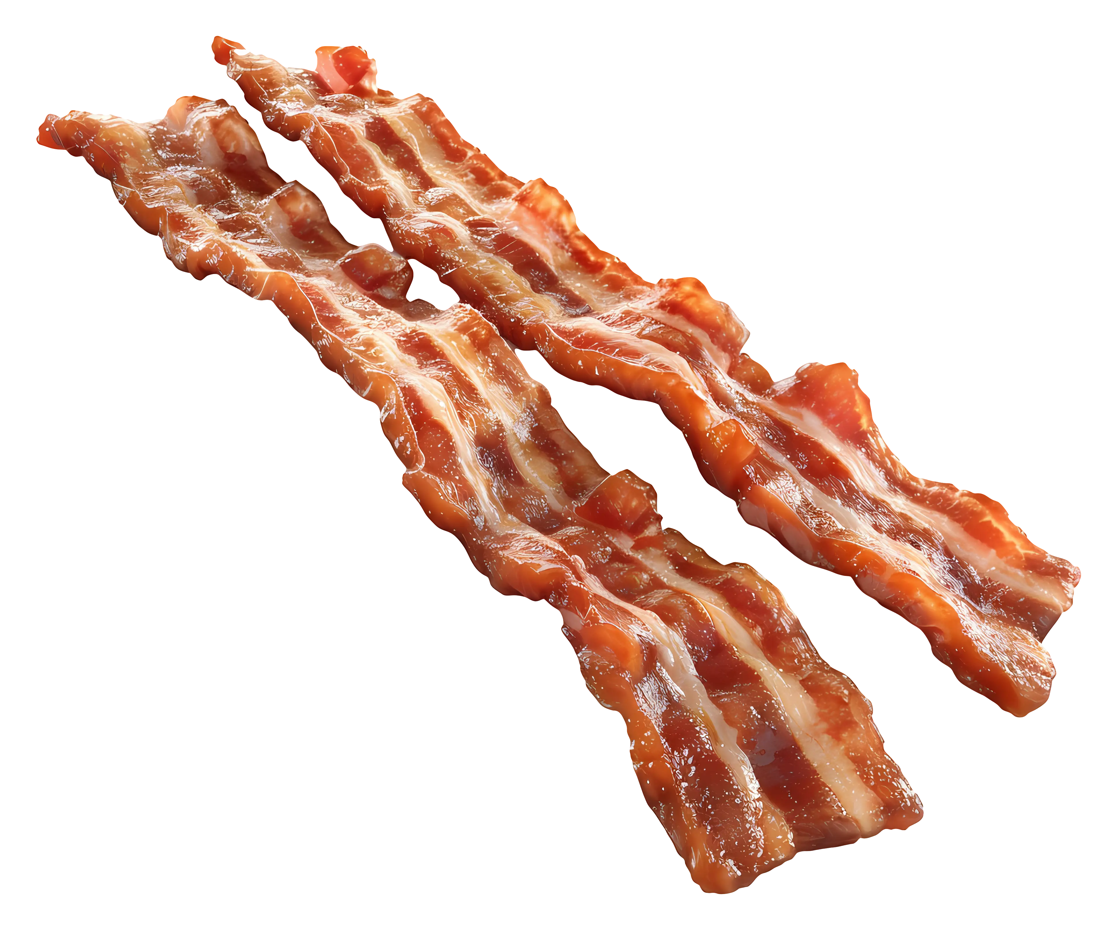
Illustration of Crispy Bacon Strips -

Red Virus Illustration Representing Pathogen -
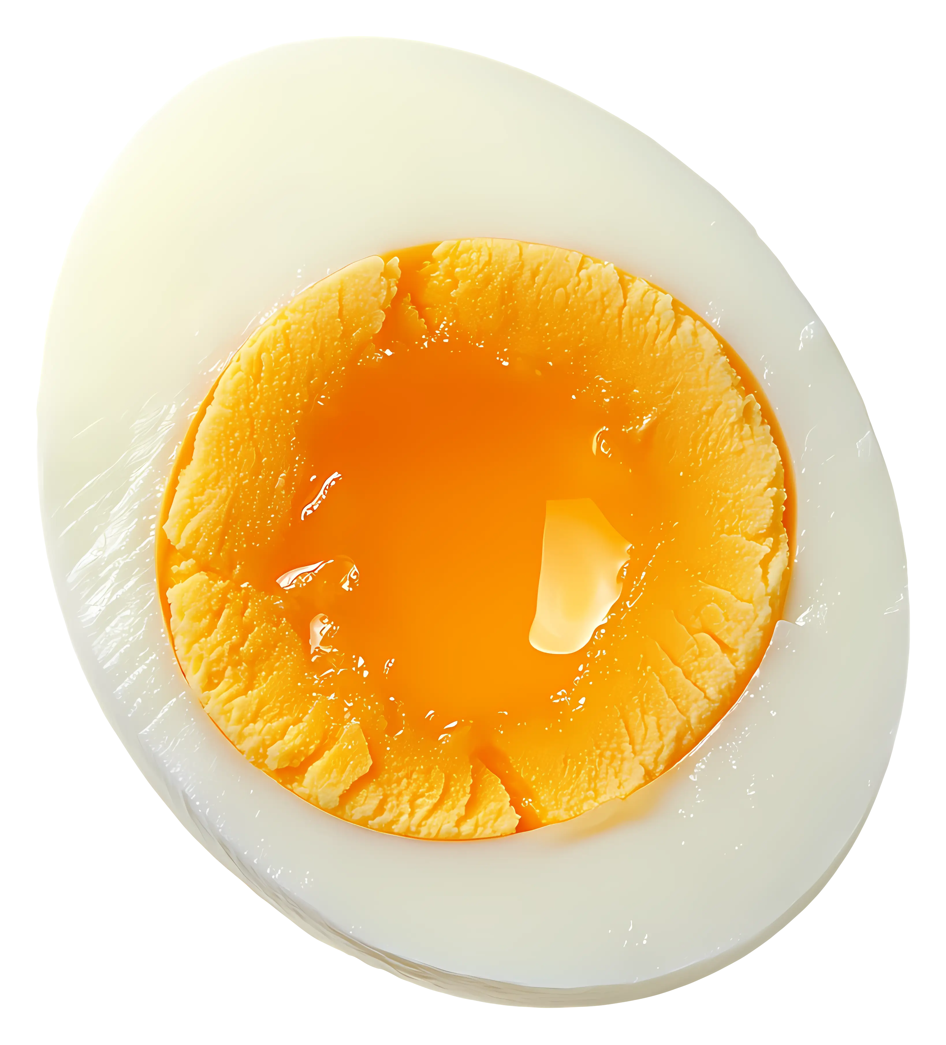
Half Boiled Egg with Soft Yolk -
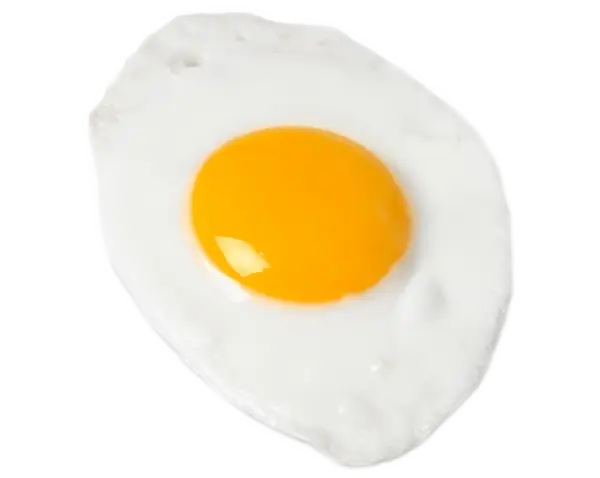
Fried Egg Illustration -
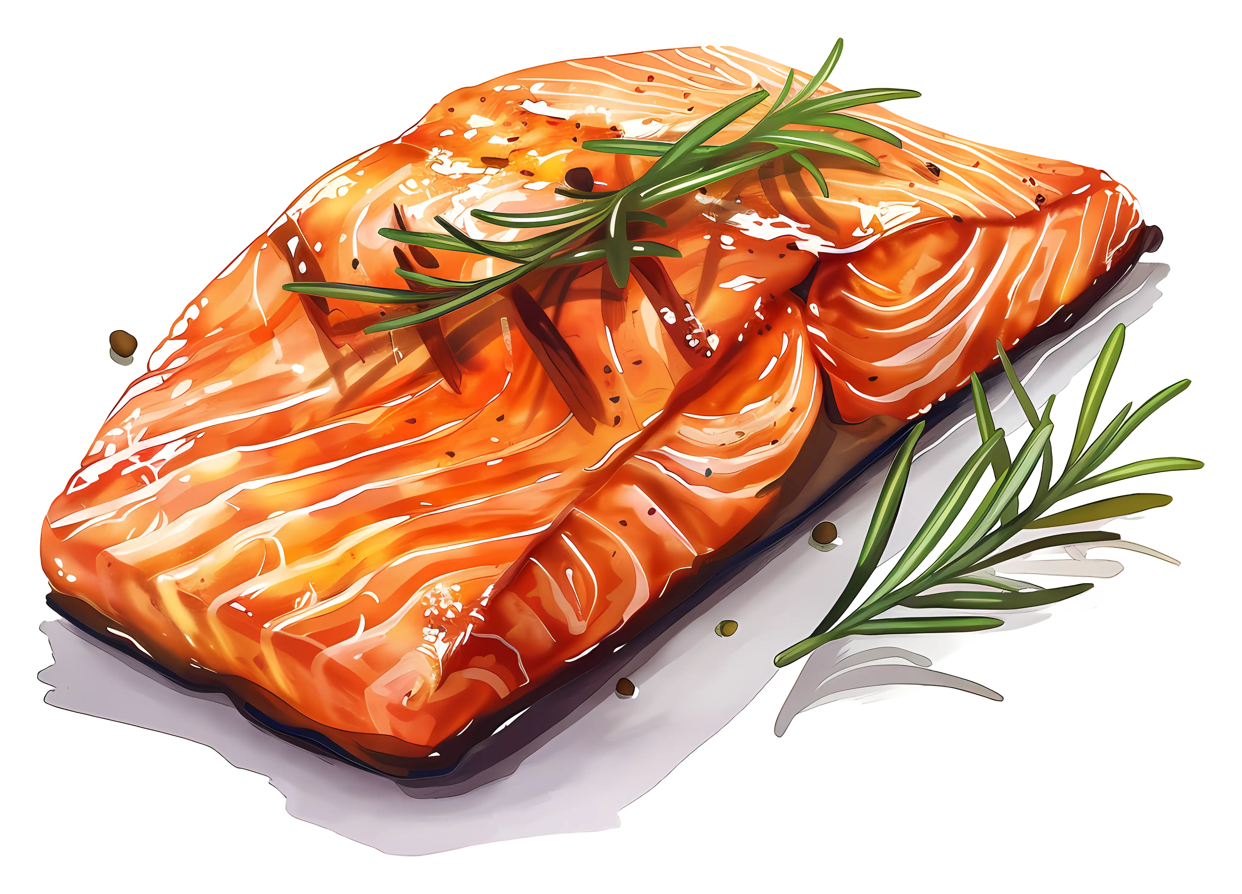
Roasted Salmon with Rosemary Garnish -

Fried Egg with Garnish Illustration -
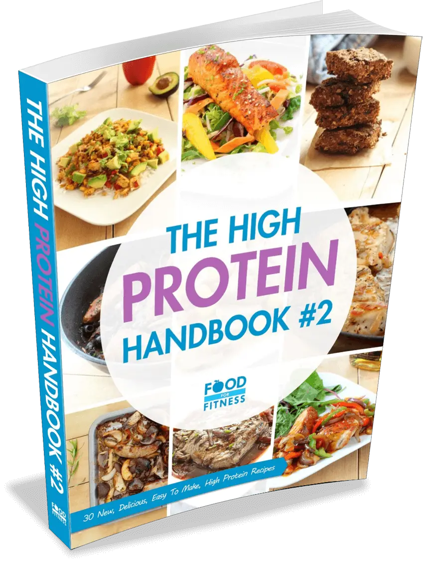
The High Protein Handbook #2 -
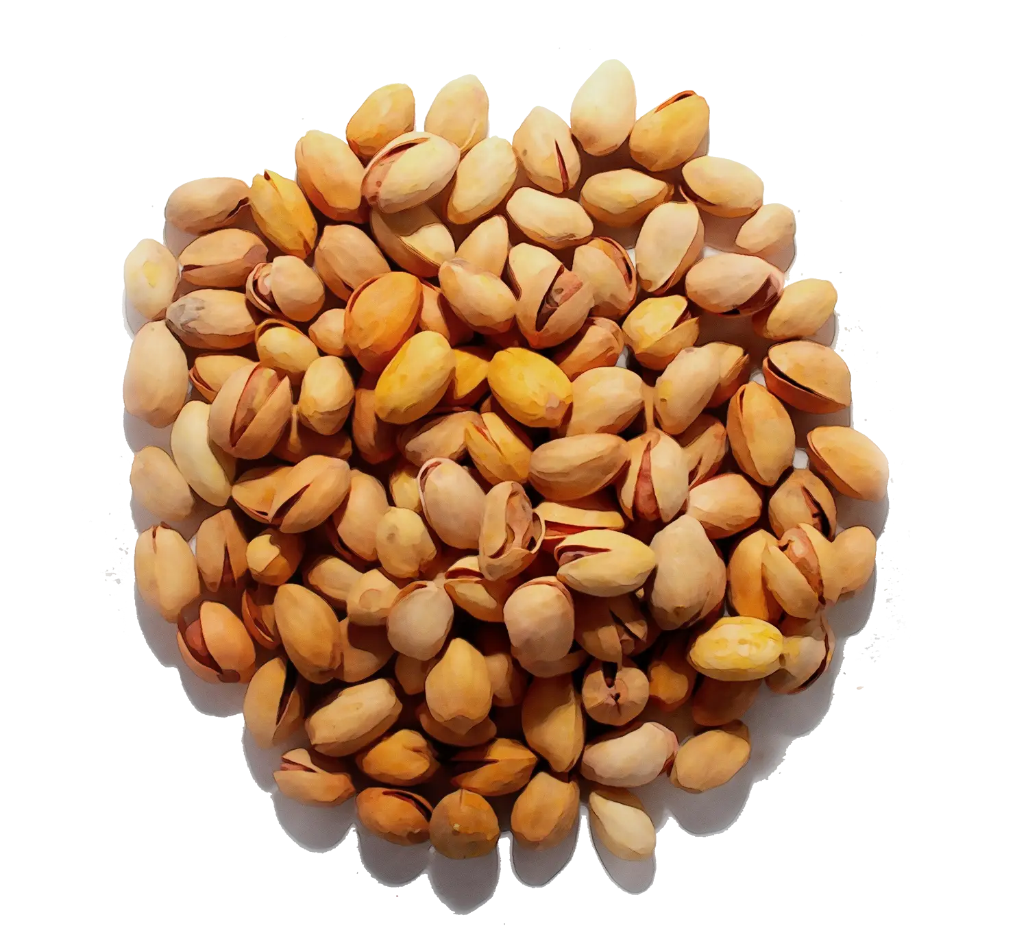
Pile of Pistachio Nuts -

Scientific Chemical Structure Diagram -
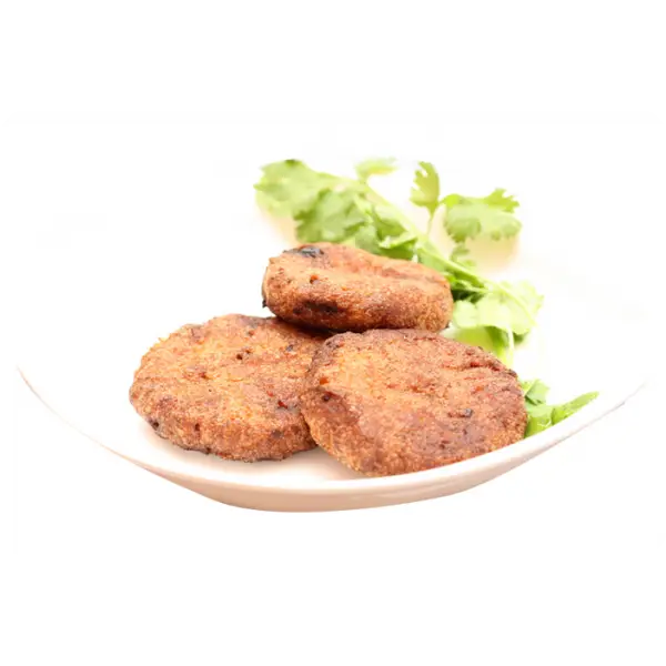
Chicken Patties with Herbs -
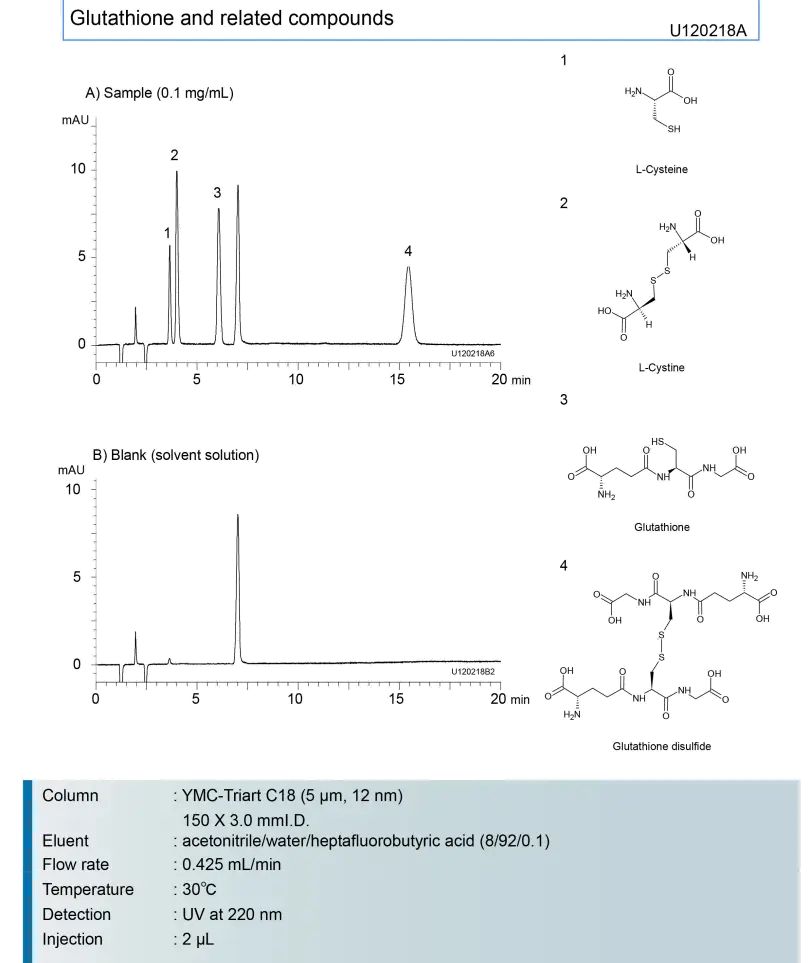
Chemical Analysis and Data Representation -
Cross Made of DNA Strands -
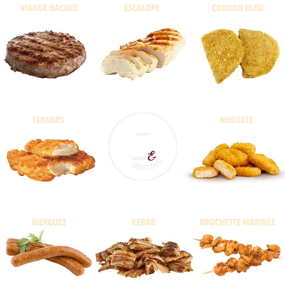
Assorted Meat Food Items -
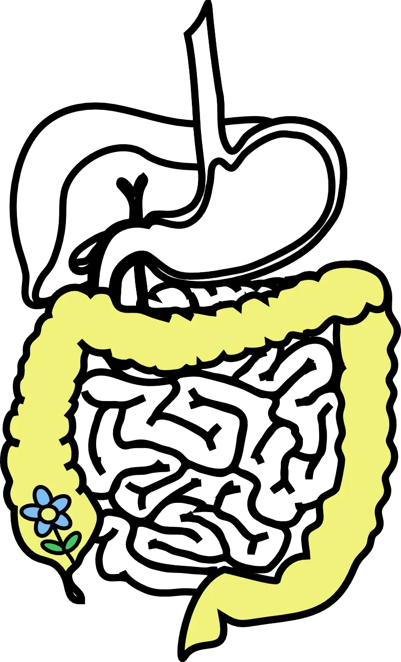
Digestive System Diagram -
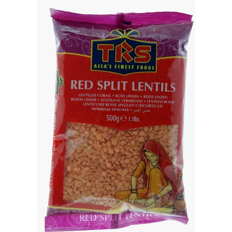
TRS Red Split Lentils Package -
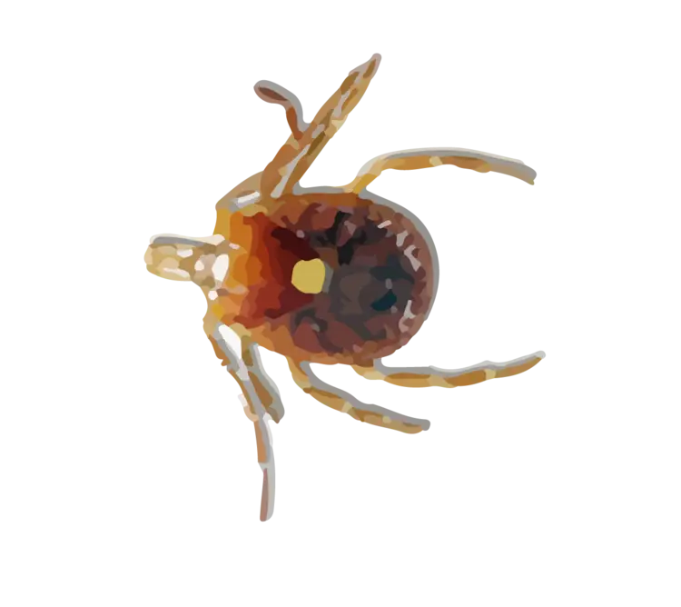
Brown Tick Close-Up Illustration -
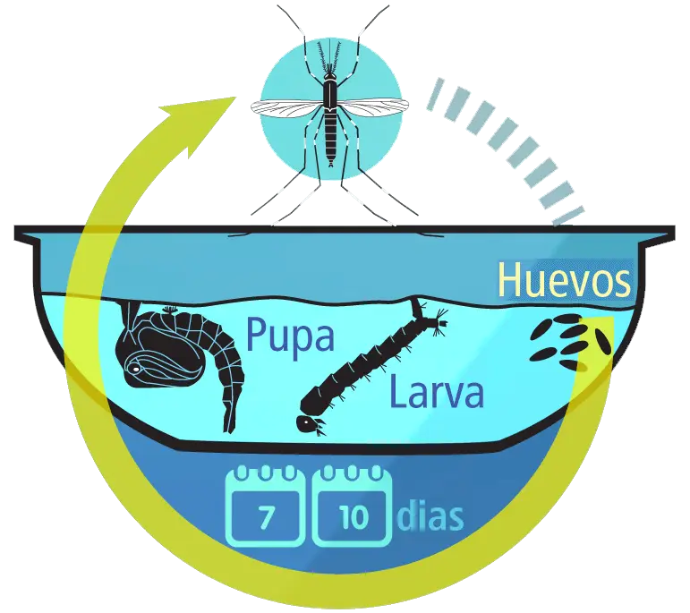
Mosquito Life Cycle Diagram -
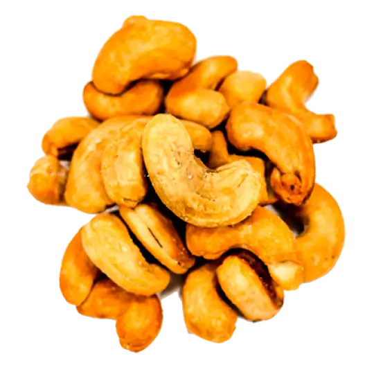
Crunchy Roasted Cashew Nuts for Healthy Snacking -
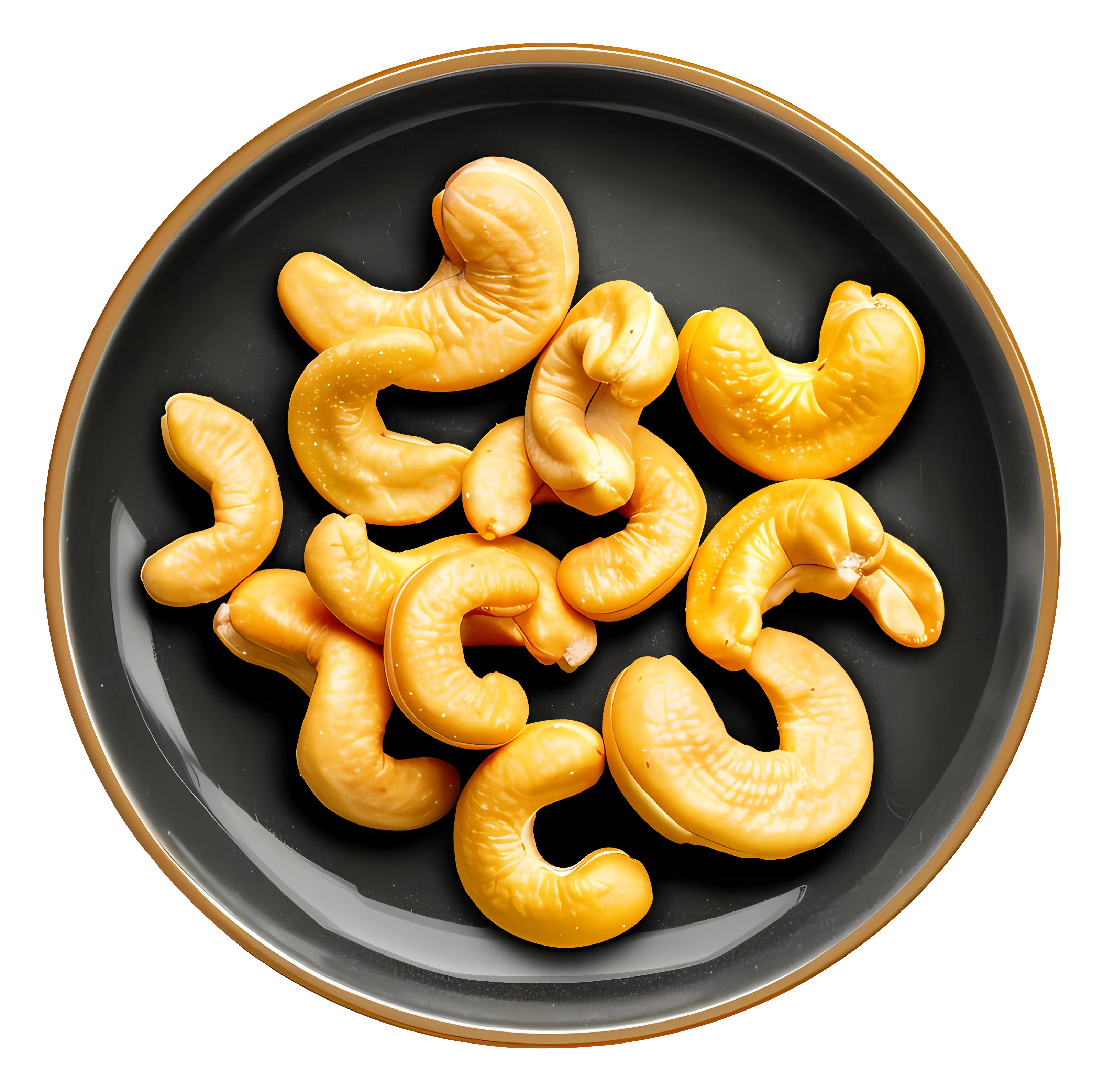
Cashew Nuts Served in a Black Bowl -
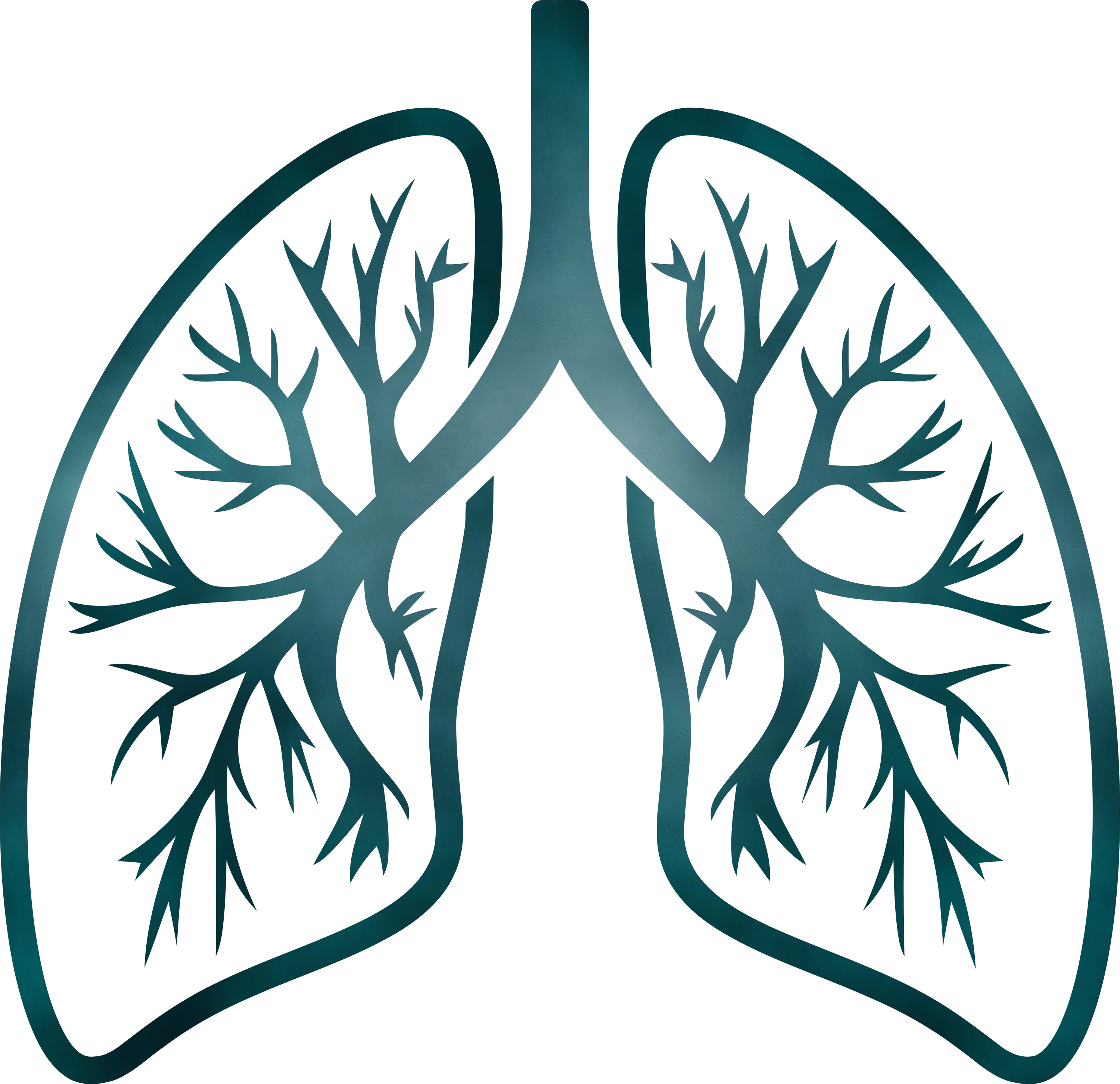
Detailed Lungs Diagram Illustration -
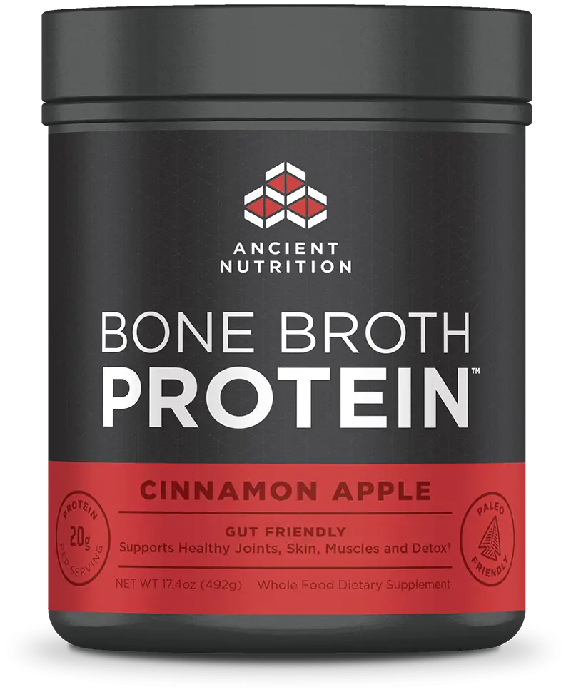
Bone Broth Protein Jar in Cinnamon Apple Flavor -
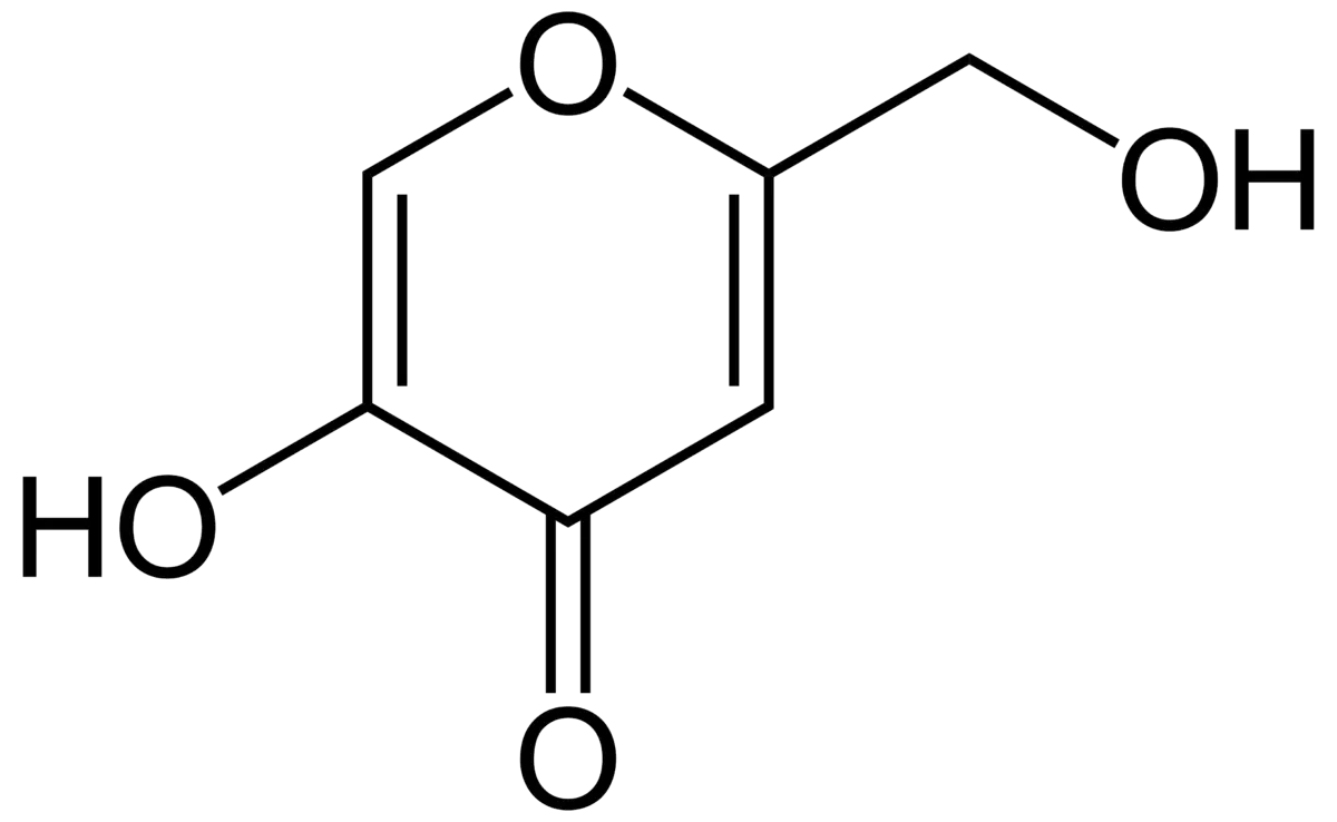
Hydroqui Organic Compound Diagram -
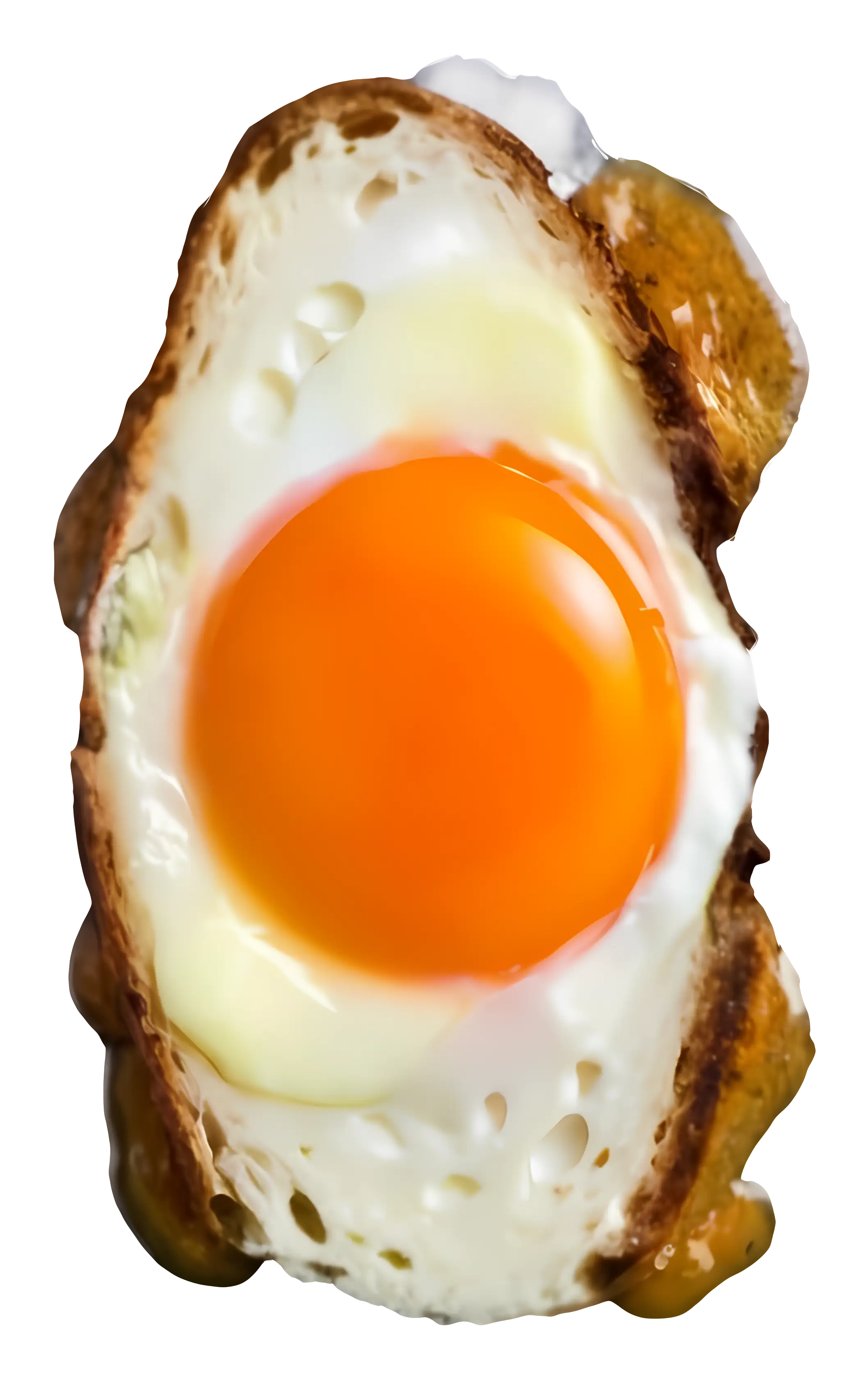
Realistic Fried Egg -

Chemical Structure Diagram -
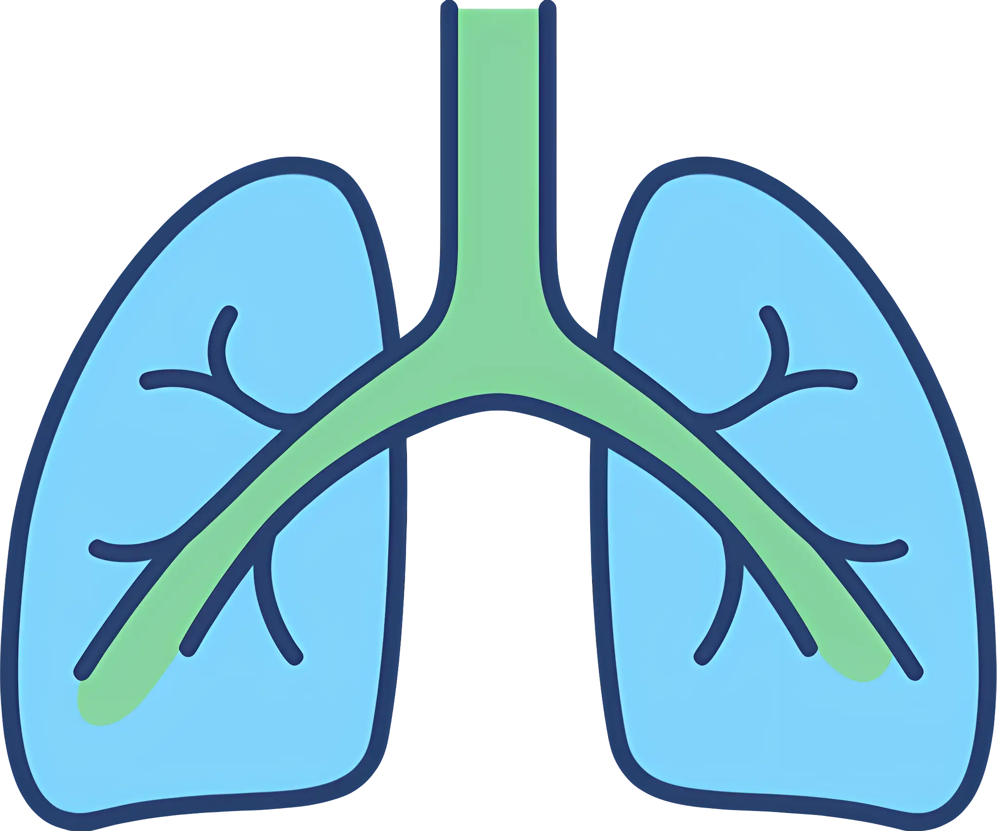
Medical Illustration of Human Lungs -
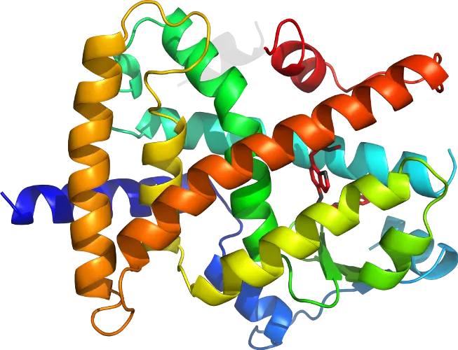
Protein Molecular Structure Model -

Boiled Egg with Soft Yolk -
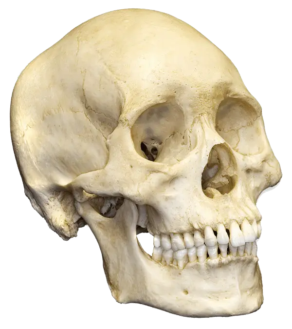
Human Skull Model for Anatomy Study -
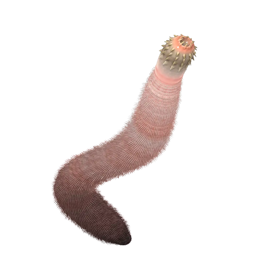
Illustration of a Worm